
















©The Author(s) 2024.
World J Radiol. Jul 28, 2024; 16(7): 265-273
Published online Jul 28, 2024. doi: 10.4329/wjr.v16.i7.265
Published online Jul 28, 2024. doi: 10.4329/wjr.v16.i7.265
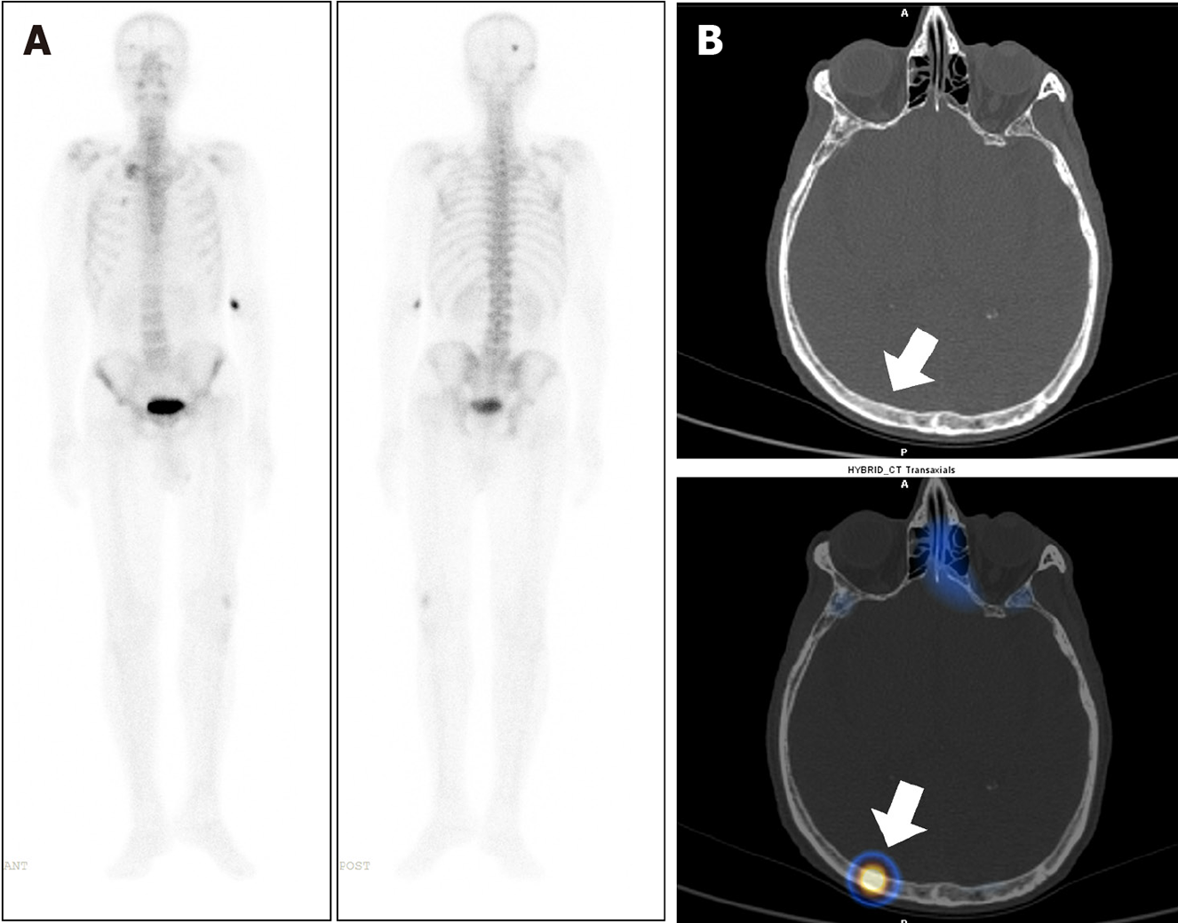
Figure 4 A 76-year-old male, with a newly diagnosed case of carcinoma of the prostate underwent whole body bone scintigraphy.
A: 99mTc methylene diphosphonate Whole body planar images show solitary focal area of increased tracer uptake involving skull bone on the right side; B: Axial computed tomography (CT) and fused single photon emission computed tomography-CT images show tracer localization to the right parietal bone (arrow) with no significant CT abnormality to suggest of metastatic disease involvement. The lesion was thus considered indeterminate.
- Citation: Singh P, Agrawal K, Rahman A, Singhal T, Parida GK, Gnanasegaran G. Incidence of exclusive extrapelvic skeletal metastasis in prostate carcinoma on bone scintigraphy. World J Radiol 2024; 16(7): 265-273
- URL: https://www.wjgnet.com/1949-8470/full/v16/i7/265.htm
- DOI: https://dx.doi.org/10.4329/wjr.v16.i7.265