
















©The Author(s) 2024.
World J Radiol. Apr 28, 2024; 16(4): 94-108
Published online Apr 28, 2024. doi: 10.4329/wjr.v16.i4.94
Published online Apr 28, 2024. doi: 10.4329/wjr.v16.i4.94
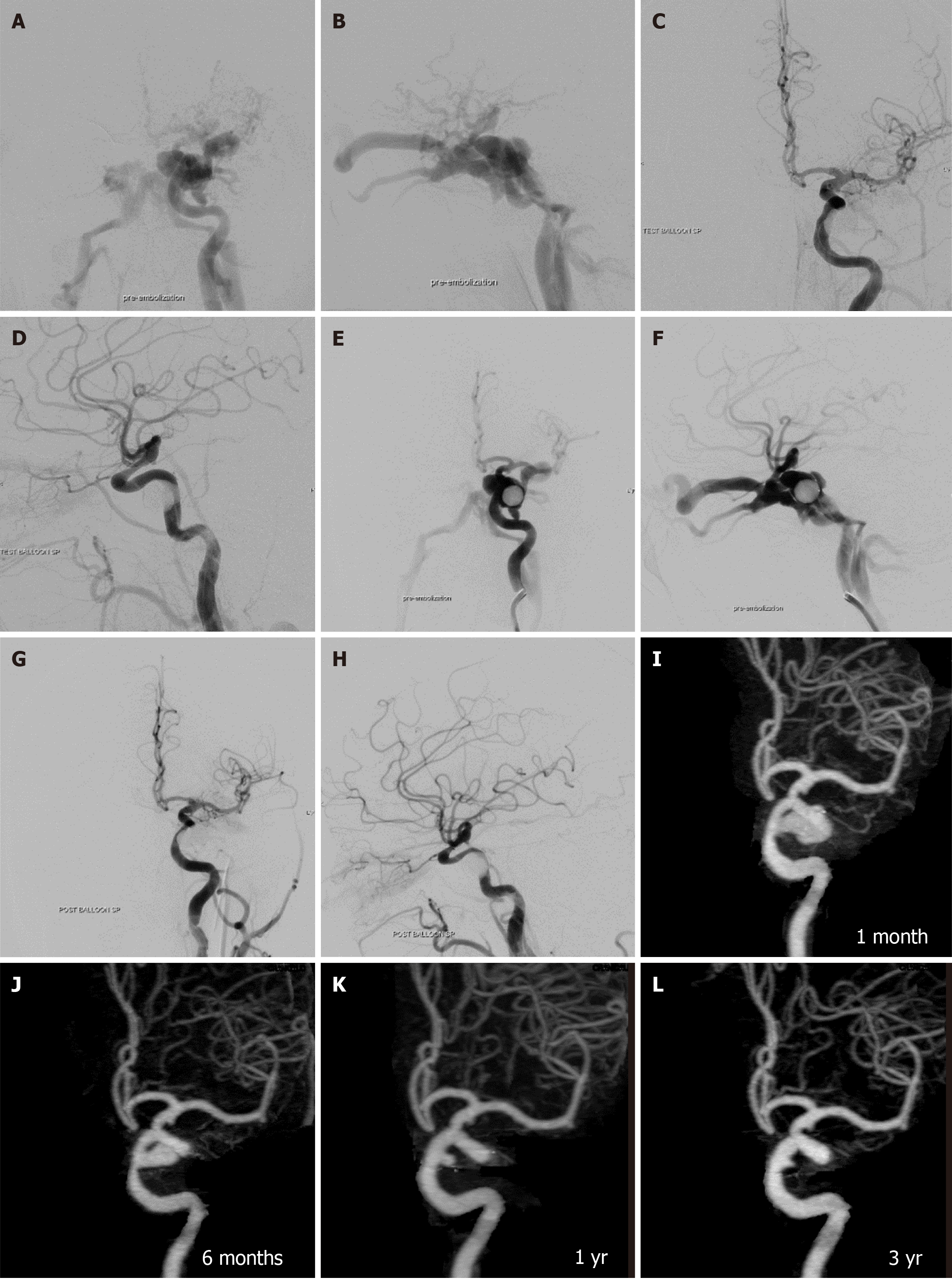
Figure 6 A 27-year-old male presented with left proptosis and red eye immediately after a motor vehicle accident.
A and B: Anteroposterior (AP) and lateral views of the left internal carotid artery (LICA) injection showed large traumatic carotid-cavernous fistula without antegrade flow into the anterior and middle cerebral arteries; C and D: AP and lateral views of the LICA revealed complete obliteration of the fistula after the detachment of the balloon; E and F: AP and lateral views of the LICA obtained 1 wk later demonstrated recurrent traumatic carotid-cavernous fistula; G and H: AP and lateral views of the LICA confirmed complete obliteration after retreatment with balloon embolization; I-L: AP views of the three-dimensional reconstructed images of the LICA using computed tomography angiography at 1 month (I), 6 months (J), 1 year (K), and 3 years (L) on the same projection demonstrated the regression of pseudoaneurysm at C5 cavernous segment of the LICA from large (grade 3) to small (grade 1) size.
- Citation: Iampreechakul P, Wangtanaphat K, Chuntaroj S, Wattanasen Y, Hangsapruek S, Lertbutsayanukul P, Puthkhao P, Siriwimonmas S. Pseudoaneurysm formation following transarterial embolization of traumatic carotid-cavernous fistula with detachable balloon: An institutional cohort long-term study. World J Radiol 2024; 16(4): 94-108
- URL: https://www.wjgnet.com/1949-8470/full/v16/i4/94.htm
- DOI: https://dx.doi.org/10.4329/wjr.v16.i4.94