
















©The Author(s) 2024.
World J Radiol. Dec 28, 2024; 16(12): 722-748
Published online Dec 28, 2024. doi: 10.4329/wjr.v16.i12.722
Published online Dec 28, 2024. doi: 10.4329/wjr.v16.i12.722
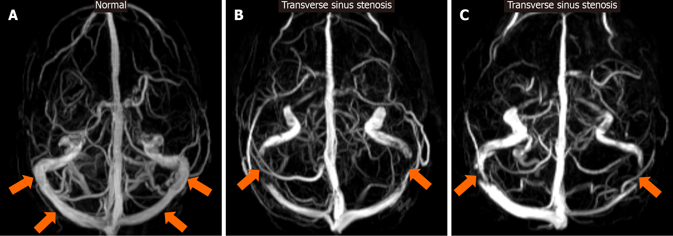
Figure 12 Transverse sinus stenosis.
A: For reference, a maximum-intensity-projection axial reconstruction of a 3D phase contrast magnetic resonance venography examination of the brain in a normal patient is provided. Note that the caliber of the transverse sinus bilaterally is within expected-normal limits (arrows); B and C: Display axial maximum-intensity-projection 3D phase contrast magnetic resonance venography reconstructions in different patients with signs and symptoms of increased intracranial pressure, demonstrating significant bilateral transverse sinus stenosis (arrows).
- Citation: Arkoudis NA, Davoutis E, Siderakis M, Papagiannopoulou G, Gouliopoulos N, Tsetsou I, Efthymiou E, Moschovaki-Zeiger O, Filippiadis D, Velonakis G. Idiopathic intracranial hypertension: Imaging and clinical fundamentals. World J Radiol 2024; 16(12): 722-748
- URL: https://www.wjgnet.com/1949-8470/full/v16/i12/722.htm
- DOI: https://dx.doi.org/10.4329/wjr.v16.i12.722