
















©The Author(s) 2024.
World J Radiol. Oct 28, 2024; 16(10): 608-615
Published online Oct 28, 2024. doi: 10.4329/wjr.v16.i10.608
Published online Oct 28, 2024. doi: 10.4329/wjr.v16.i10.608
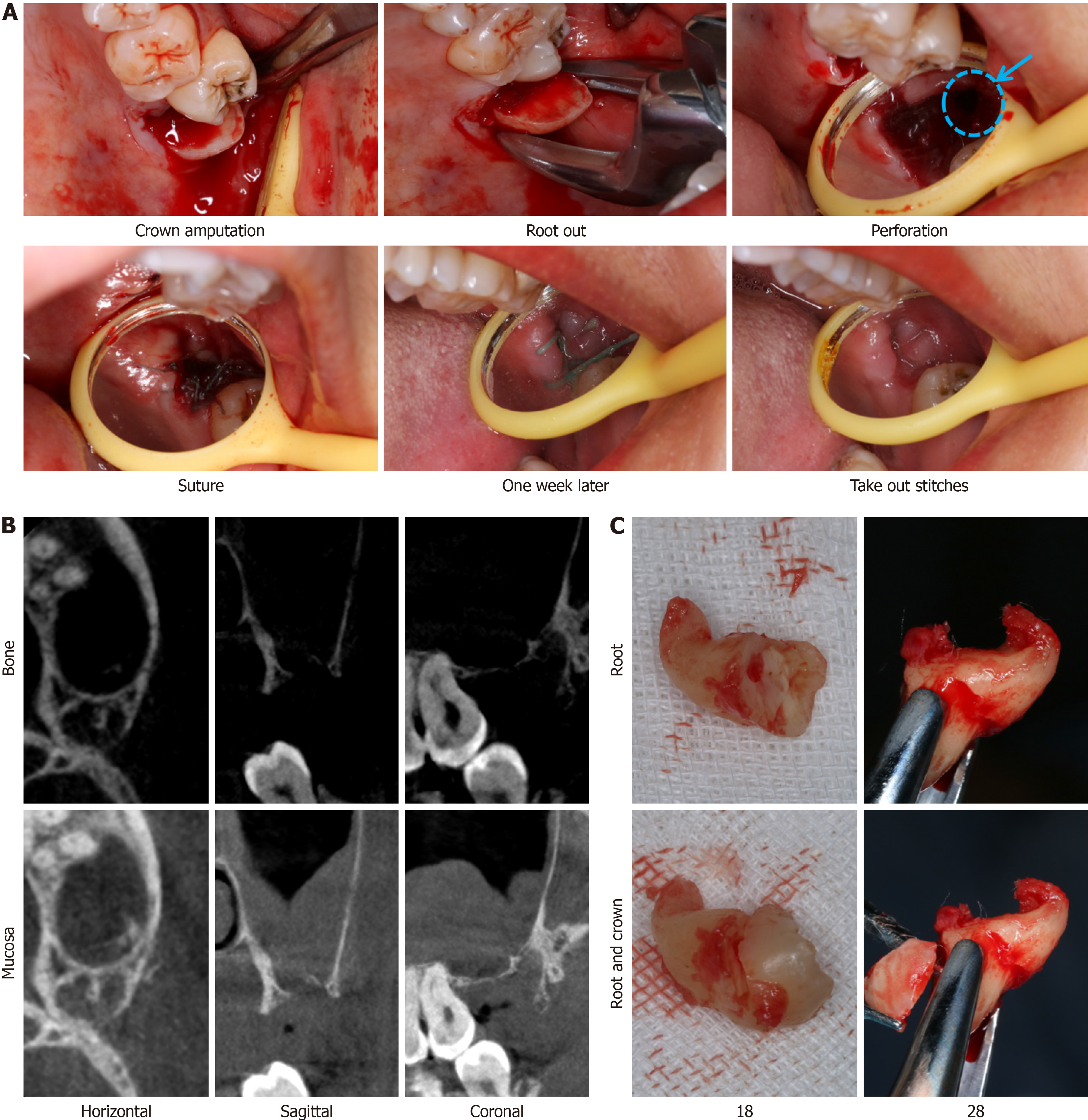
Figure 3 Intraoperative and postoperative imaging.
A: Intraoral image showing a maxillary sinus perforation (blue circle); B: Radiographs obtained two weeks postoperatively; C: Extracted tooth showing the junction of the root and crown.
- Citation: Liu H, Wang F, Tang YL, Yan X. Asymmetric outcomes in bilateral maxillary impacted tooth extractions: A case report. World J Radiol 2024; 16(10): 608-615
- URL: https://www.wjgnet.com/1949-8470/full/v16/i10/608.htm
- DOI: https://dx.doi.org/10.4329/wjr.v16.i10.608