Copyright
©The Author(s) 2023.
World J Radiol. Apr 28, 2023; 15(4): 98-117
Published online Apr 28, 2023. doi: 10.4329/wjr.v15.i4.98
Published online Apr 28, 2023. doi: 10.4329/wjr.v15.i4.98
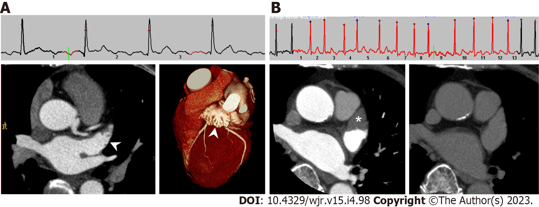
Figure 1 Cardiac computed tomography images of left atrial appendage in patients with sinus rhythm and atrial fibrillation.
A: Under normal sinus rhythm, left atrial appendage (LAA) is filled with contrast medium in early phase, which evaluates the coronary artery. Left: axial image; right: three-dimensional volume-rendered image; B: Under atrial fibrillation, axial early phase image shows triangular filling defect in LAA (left, asterisk). On delayed phase (right), LAA filling defect completely disappeared, which means the filling defect reflected blood stasis in LAA rather than thrombus.
- Citation: Yoshihara S. Evaluation of causal heart diseases in cardioembolic stroke by cardiac computed tomography. World J Radiol 2023; 15(4): 98-117
- URL: https://www.wjgnet.com/1949-8470/full/v15/i4/98.htm
- DOI: https://dx.doi.org/10.4329/wjr.v15.i4.98


