Copyright
©The Author(s) 2023.
World J Radiol. Apr 28, 2023; 15(4): 127-135
Published online Apr 28, 2023. doi: 10.4329/wjr.v15.i4.127
Published online Apr 28, 2023. doi: 10.4329/wjr.v15.i4.127
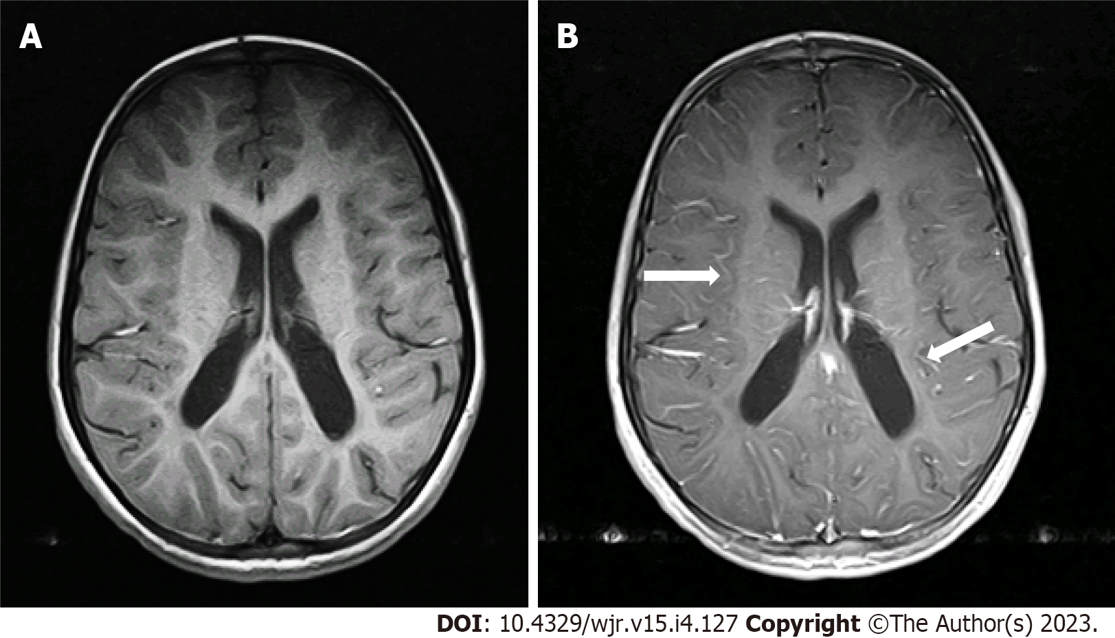
Figure 2 Moderate pseudo leptomeningeal contrast enhancement a 7-yr-old female with developmental delays (Grade 2).
A: Axial spin echo T1-weighted imaging; B: Axial post-contrast T1-weighted imaging showed pseudo leptomeningeal contrast enhancement as smooth and slightly thickened enhancements (arrows) throughout the depths of the sulci.
- Citation: Hilal K, Khandwala K, Rashid S, Khan F, Anwar SSM. Does sevoflurane sedation in pediatric patients lead to “pseudo” leptomeningeal enhancement in the brain on 3 Tesla magnetic resonance imaging? World J Radiol 2023; 15(4): 127-135
- URL: https://www.wjgnet.com/1949-8470/full/v15/i4/127.htm
- DOI: https://dx.doi.org/10.4329/wjr.v15.i4.127


