Copyright
©The Author(s) 2023.
World J Radiol. Feb 28, 2023; 15(2): 42-55
Published online Feb 28, 2023. doi: 10.4329/wjr.v15.i2.42
Published online Feb 28, 2023. doi: 10.4329/wjr.v15.i2.42
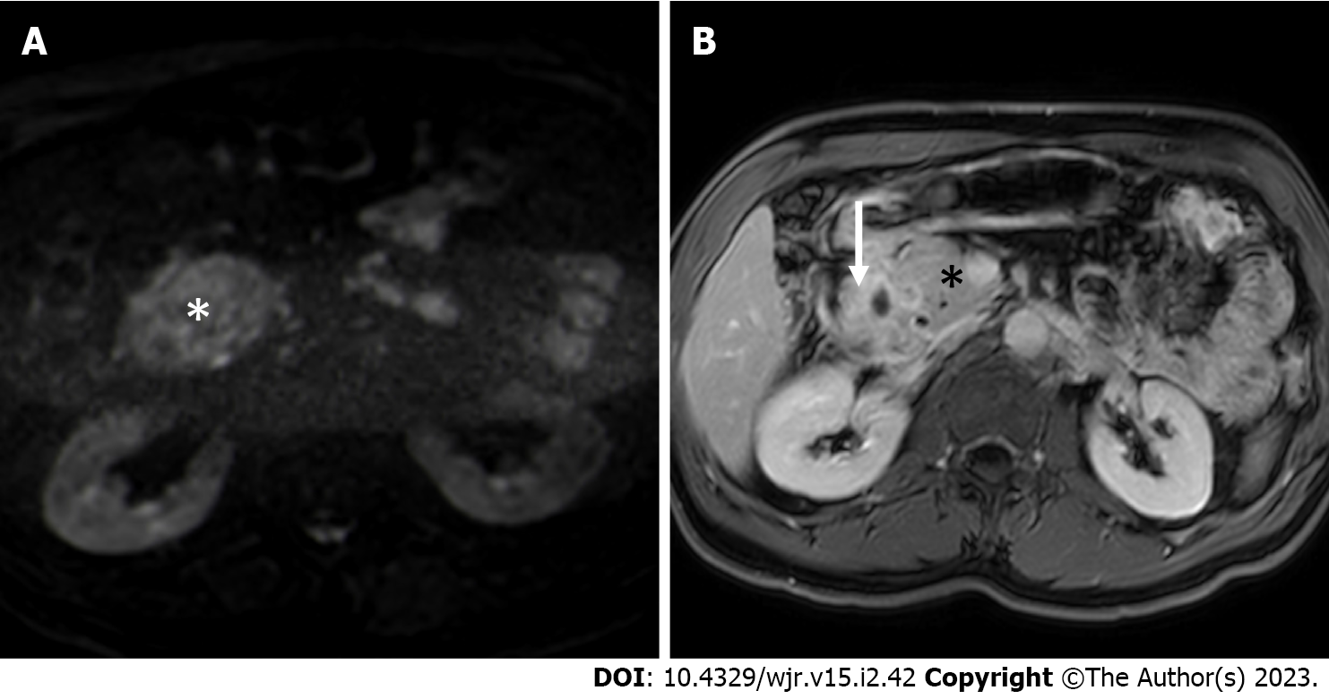
Figure 4 Magnetic resonance imaging.
A: Axial high b value (b = 800 s/mm2) diffusion weighted imaging image shows absence of increased diffusivity restriction in the thickened groove area (star) in comparison to adjacent “normal” pancreas, finding associated with paraduodenal pancreatitis and uncommon in pancreatic cancer; B: Axial delayed phase T1-weighted magnetic resonance imaging acquisition shows increased enhancement of the duodenal walls and of the groove region (arrow) in comparison to “normal” pancreas (star), finding often associated with paraduodenal pancreatitis.
- Citation: Bonatti M, De Pretis N, Zamboni GA, Brillo A, Crinò SF, Valletta R, Lombardo F, Mansueto G, Frulloni L. Imaging of paraduodenal pancreatitis: A systematic review. World J Radiol 2023; 15(2): 42-55
- URL: https://www.wjgnet.com/1949-8470/full/v15/i2/42.htm
- DOI: https://dx.doi.org/10.4329/wjr.v15.i2.42


