Copyright
©The Author(s) 2022.
World J Radiol. Sep 28, 2022; 14(9): 329-341
Published online Sep 28, 2022. doi: 10.4329/wjr.v14.i9.329
Published online Sep 28, 2022. doi: 10.4329/wjr.v14.i9.329
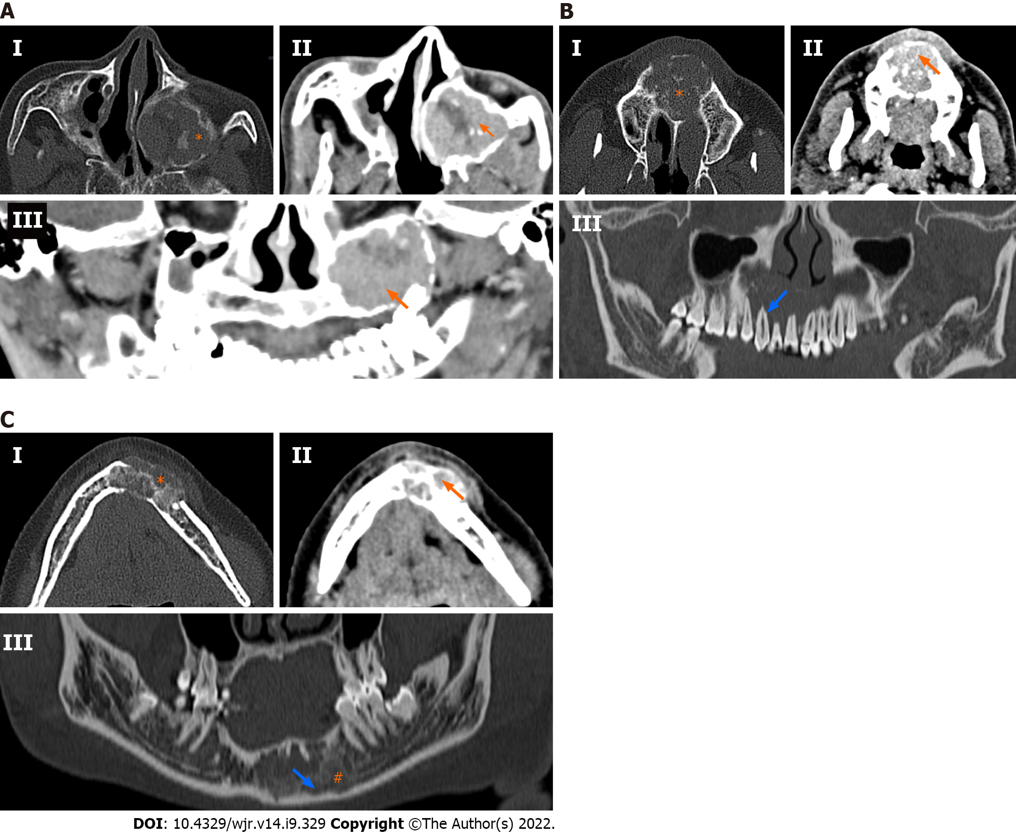
Figure 2 Spectrum of multidetector computed tomography findings in central giant cell granulomas.
A: 35-year-old woman presented with upper facial pain and nasal obstruction. Cone beam computed tomography (CBCT) shows a left-sided unilocular lytic lesion arising from the left maxilla (Panel I: Bone window) with compression of the maxillary sinus. Mineralised matrix was scattered in the substance of the tumour (asterisk). The lesion showed a significant soft tissue component, which enhanced to an extent greater than the surrounding muscles [arrow, Panel II and III: Axial and curved multiplanar reconstructed (MPR) coronal soft tissue images]. Hyperenhancement of the soft tissue tumour component was highly suggestive of a prospective central giant cell granuloma (CGCG) diagnosis; B: A 30-year-old man presented with pain and upper jaw swelling, contrast-enhanced computed tomography (CECT) showed a lytic sclerotic, multilocular mass arising from the maxilla with the presence of incomplete septae (asterisk) with mineralised matrix (Panel I: Axial bone window). Significant solid soft tissue component with enhancement greater (arrow) than the surrounding muscles was also noted (Panel II: Axial soft tissue window images). Curved MPR images (Panel III: Bone window) showed resorption of the roots (empty arrow) and floor of the nasal cavity; C: A 24-year-old woman presented with progressive jaw swelling over the last 6 mo, with intermittent pain. CECT showed a sclerotic lytic lesion with a honeycomb appearance (Panel I: Axial bone window) arising from the mandible. The lesion showed thick bony septae with mineralised matrix (asterisk). The associated soft tissue component showed enhancement similar to the surrounding muscles (orange arrow: Panel II: Axial soft tissue window). The tumour (blue arrow) can be seen encroaching onto the distal end (#) of the left inferior alveolar canal (Panel III: Curved MPR bone window).
- Citation: Ghosh A, Lakshmanan M, Manchanda S, Bhalla AS, Kumar P, Bhutia O, Mridha AR. Contrast-enhanced multidetector computed tomography features and histogram analysis can differentiate ameloblastomas from central giant cell granulomas . World J Radiol 2022; 14(9): 329-341
- URL: https://www.wjgnet.com/1949-8470/full/v14/i9/329.htm
- DOI: https://dx.doi.org/10.4329/wjr.v14.i9.329


