Copyright
©The Author(s) 2022.
World J Radiol. Aug 28, 2022; 14(8): 256-271
Published online Aug 28, 2022. doi: 10.4329/wjr.v14.i8.256
Published online Aug 28, 2022. doi: 10.4329/wjr.v14.i8.256
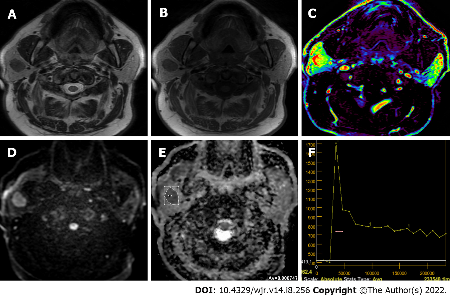
Figure 2 Sixty-five years old male patient with smooth lobule contoured Warthin’s tumor located on the superficial lobe of right parotid gland.
A and B: Hypointense signal of the lesion compared to the gland on T2-weighted image and T1-weighted image; C: The mass is hyperperfused on the color-coded perfusion image; D: The mass appears to be slightly heterogenous hyperintense on the diffusion-weighted image, E: The apparent diffusion coefficient (ADC) value of mass was 0.74 × 10⁻³ mm2/s on the ADC map; F: The time intensity curve of mass has a wash-out ratio of 50%.
- Citation: Gökçe E, Beyhan M. Advanced magnetic resonance imaging findings in salivary gland tumors. World J Radiol 2022; 14(8): 256-271
- URL: https://www.wjgnet.com/1949-8470/full/v14/i8/256.htm
- DOI: https://dx.doi.org/10.4329/wjr.v14.i8.256


