
















©The Author(s) 2022.
World J Radiol. Feb 28, 2022; 14(2): 30-46
Published online Feb 28, 2022. doi: 10.4329/wjr.v14.i2.30
Published online Feb 28, 2022. doi: 10.4329/wjr.v14.i2.30
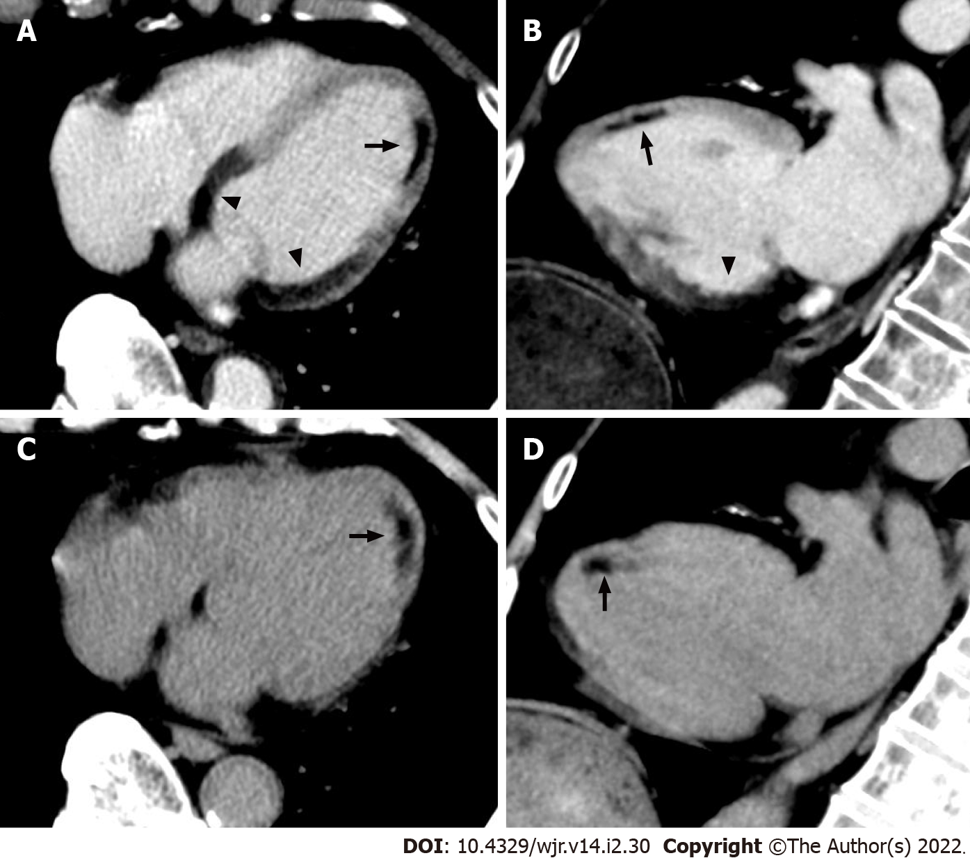
Figure 17 Non-electrocardiogram-gated contrast-enhanced computed tomography images in acute coronary syndrome of right coronary artery complicated with anterior old myocardial infarction.
An 82-year-old man with back pain underwent non-electrocardiogram (ECG)-gated contrast-enhanced computed tomography (CECT) in search of aortic dissection. Axial (A) and vertical long axis (B) reformatted non-ECG-gated CECT images acquired 120 s after contrast injection showed decreased myocardial enhancement in the basal to mid inferior, inferolateral, and inferoseptal walls of the left ventricle (arrowheads). Non-ECG-gated CECT images also showed decreased myocardial enhancement in the mid anterior wall of the left ventricle apart from the above-mentioned area (arrow). Axial (C) and vertical long axis (D) reformatted non-ECG-gated non-contrast CT images showed subendocardial low attenuation less than 0 Hounsfield units only in the mid anterior wall of the left ventricle, which is consistent with myocardial fatty degeneration associated with the old myocardial infarction (arrow).
- Citation: Yoshihara S. Acute coronary syndrome on non-electrocardiogram-gated contrast-enhanced computed tomography. World J Radiol 2022; 14(2): 30-46
- URL: https://www.wjgnet.com/1949-8470/full/v14/i2/30.htm
- DOI: https://dx.doi.org/10.4329/wjr.v14.i2.30