Copyright
©The Author(s) 2021.
World J Radiol. Sep 28, 2021; 13(9): 258-282
Published online Sep 28, 2021. doi: 10.4329/wjr.v13.i9.258
Published online Sep 28, 2021. doi: 10.4329/wjr.v13.i9.258
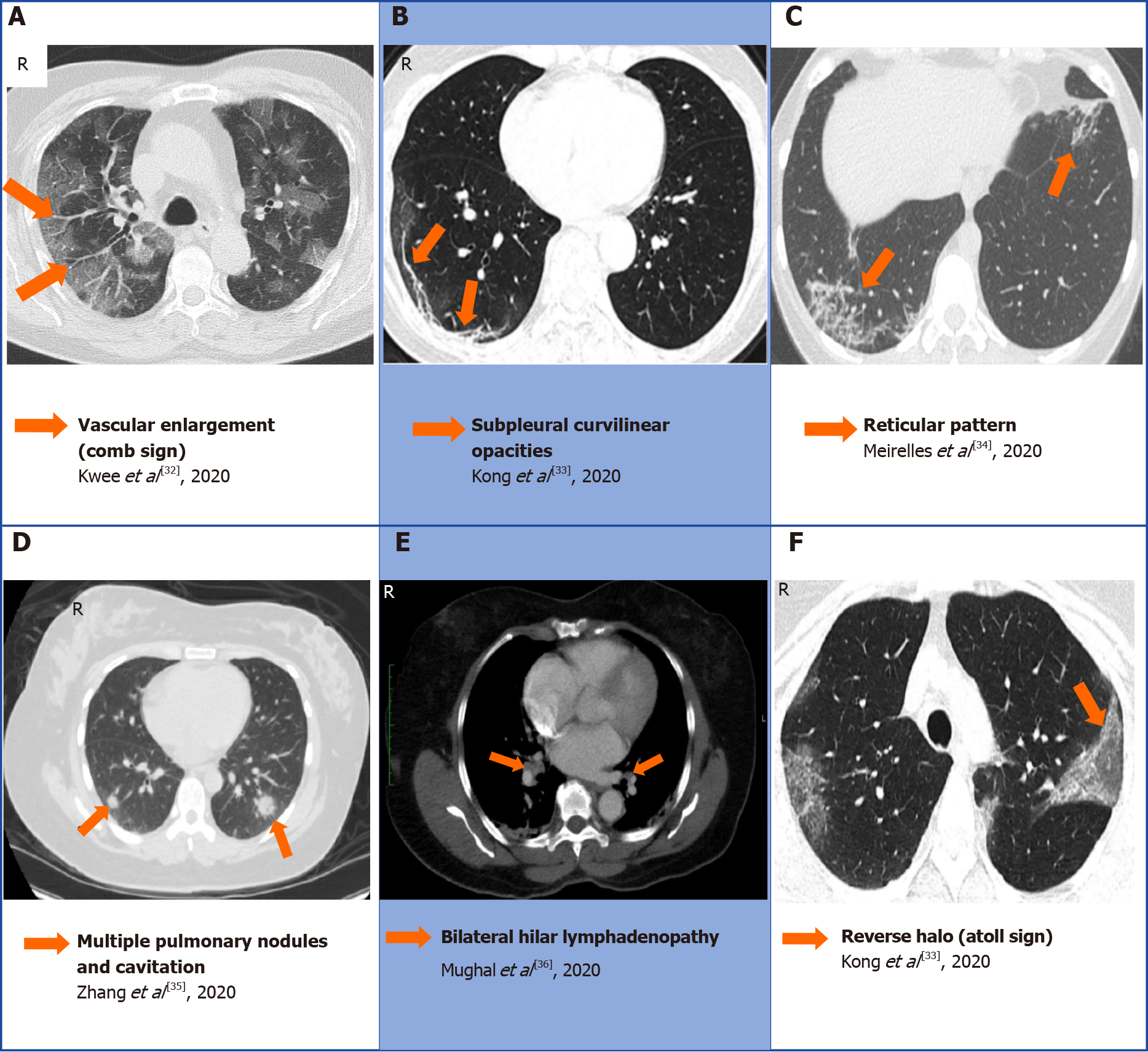
Figure 7 A collection of chest computed tomography that displays some of the atypical findings of coronavirus disease 2019 pneumonia[32-36].
A: Comb sign in the right lobe characterized by vascular enlargement; B: Curvilinear opacities in the subpleural area; C: Reticular pattern bilaterally; D: Multiple nodules and cavitation; E: Bilateral hilar lymphadenopathy; F: Atoll sign also known as reverse halo. A: Citation: Kwee TC, Kwee RM. Chest CT in COVID-19: What the radiologist needs to know. Radiographics 2020; 40: 1848-1865. Copyright ©The Author(s) 2021. Published by Radiographics; B and F: Citation: Kong W, Agarwal PP. Chest imaging appearance of COVID-19 infection. Radiology: Cardiothoracic Imaging 2020; 2: e200028. Copyright ©The Author(s) 2020. Published by the Radiological Society of North America, Inc; C: Citation: Meirelles GSP. COVID-19: A brief update for radiologists. Radiologia Brasileira 2020; 53: 320-328. Copyright ©The Author(s) 2020. Published by Radiology brasil; D: Citation: Zhang Q, Douglas A, Abideen ZU, Khanal S, Tzarnas S. Novel coronavirus (2019-nCoV) in disguise. Cureus 2020; 12: e7521. Copyright ©The Author(s) 2020. Published by Cureus; E: Citation: Mughal MS, Rehman R, Osman R, Kan N, Mirza H, Eng MH. Hilar lymphadenopathy, a novel finding in the setting of coronavirus disease (COVID-19): A case report. Journal of Medical Case Reports 2020; 14: 124. Copyright ©The Author(s) 2020. Published by BMC.
- Citation: Pal A, Ali A, Young TR, Oostenbrink J, Prabhakar A, Prabhakar A, Deacon N, Arnold A, Eltayeb A, Yap C, Young DM, Tang A, Lakshmanan S, Lim YY, Pokarowski M, Kakodkar P. Comprehensive literature review on the radiographic findings, imaging modalities, and the role of radiology in the COVID-19 pandemic. World J Radiol 2021; 13(9): 258-282
- URL: https://www.wjgnet.com/1949-8470/full/v13/i9/258.htm
- DOI: https://dx.doi.org/10.4329/wjr.v13.i9.258


