Copyright
©The Author(s) 2021.
World J Radiol. Sep 28, 2021; 13(9): 258-282
Published online Sep 28, 2021. doi: 10.4329/wjr.v13.i9.258
Published online Sep 28, 2021. doi: 10.4329/wjr.v13.i9.258
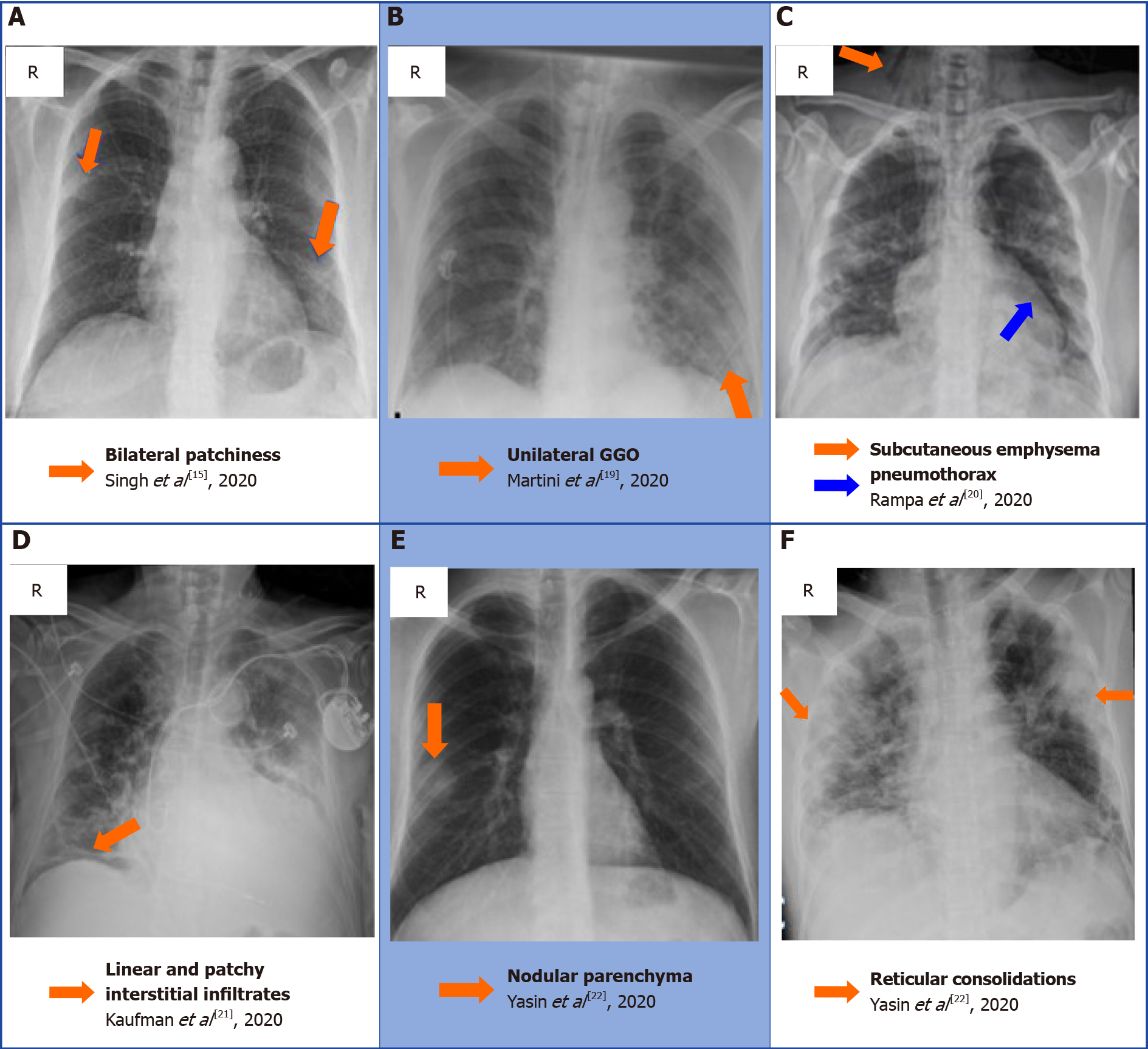
Figure 2 A collection of chest radiographs that displays some of the common and rare findings of coronavirus disease 2019 pneumonia[15,19-22].
A: Bilateral patchiness; B: Unilateral ground glass opacification; C: Subcutaneous emphysema secondary to a pneumothorax; D: Linear and patchy interstitial infiltrate in the right basal zone; E: Nodular appearance of the right lobe parenchyma; F: reticular appearance of the consolidation bilaterally. A: Citation: Singh B, Kaur P, Reid RJ, Shamoon F, Bikkina M. COVID-19 and Influenza Co-Infection: Report of Three Cases. Cureus 2020; 12: e9852. Copyright ©The Author(s) 2020. Published by Cureus; B: Citation: Martini K, Blüthgen C, Walter JE, Messerli M, Nguyen-Kim TD, Frauenfelder T. Accuracy of Conventional and Machine Learning Enhanced Chest Radiography for the Assessment of COVID-19 Pneumonia: Intra-Individual Comparison with CT. Journal of Clinical Medicine 2020;.9: 3576 Copyright ©The Author(s) 2020. Published by MDPI, Basel, Switzerland; C: Citation: Rampa L, Miceli A, Casilli F, Biraghi T, Barbara B, Donatelli F. Lung complication in COVID-19 convalescence: A spontaneous pneumothorax and pneumatocele case report. Journal of Respiratory Diseases and Medicine 2020; 2. Copyright ©The Author(s) 2020. Published by Open-access article; D: Citation: Kaufman A, Naidu S, Ramachandran S, Kaufman D, Fayad Z, Mani V. Review of radiographic findings in COVID-19. World Journal of Radiology 2020; 12: 142-55. Copyright ©The Author(s) 2020. Published by Baishideng Publishing Group Inc; E and F: Citation: Yasin R, Gouda W. Chest X-ray findings monitoring COVID-19 disease course and severity. The Egyptian Journal of Radiology and Nuclear Medicine 2020; 51: 193. Copyright ©The Author(s) 2020. Published by BMJ.
- Citation: Pal A, Ali A, Young TR, Oostenbrink J, Prabhakar A, Prabhakar A, Deacon N, Arnold A, Eltayeb A, Yap C, Young DM, Tang A, Lakshmanan S, Lim YY, Pokarowski M, Kakodkar P. Comprehensive literature review on the radiographic findings, imaging modalities, and the role of radiology in the COVID-19 pandemic. World J Radiol 2021; 13(9): 258-282
- URL: https://www.wjgnet.com/1949-8470/full/v13/i9/258.htm
- DOI: https://dx.doi.org/10.4329/wjr.v13.i9.258


