Copyright
©The Author(s) 2021.
World J Radiol. Jun 28, 2021; 13(6): 157-170
Published online Jun 28, 2021. doi: 10.4329/wjr.v13.i6.157
Published online Jun 28, 2021. doi: 10.4329/wjr.v13.i6.157
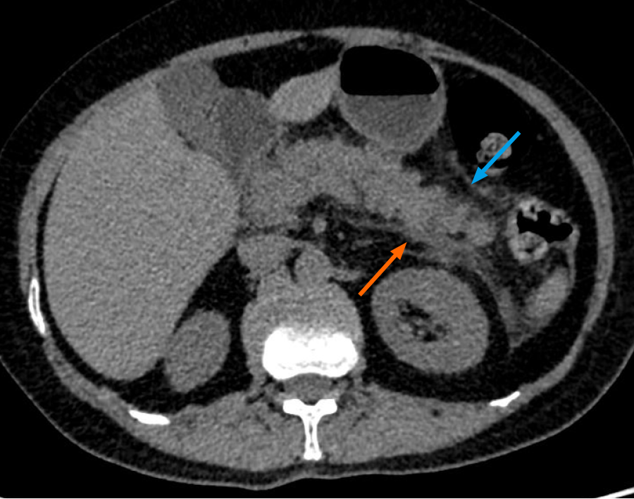
Figure 9 Acute viral pancreatitis in a coronavirus disease 2019 patient presenting with abdominal pain.
Non-contrast axial computed tomography image of the abdomen in a case of suspected viral pancreatitis (intravenous contrast could not be administered due to a history of renal parenchymal disease with elevated creatinine) is shown. The distal body and tail of pancreas reveal fuzzy margins with peri-pancreatic fat stranding (blue arrow). Thickening of the left anterior conal fascia is noted with a streak of fluid in the left retro-mesenteric plane (orange arrow). Elevated serum amylase and lipase levels, in conjunction with these imaging findings, were highly suggestive of a diagnosis of acute viral pancreatitis in a patient with coronavirus disease 2019 presenting with abdominal pain.
- Citation: Vaidya T, Nanivadekar A, Patel R. Imaging spectrum of abdominal manifestations of COVID-19. World J Radiol 2021; 13(6): 157-170
- URL: https://www.wjgnet.com/1949-8470/full/v13/i6/157.htm
- DOI: https://dx.doi.org/10.4329/wjr.v13.i6.157


