
















©The Author(s) 2021.
World J Radiol. Jun 28, 2021; 13(6): 157-170
Published online Jun 28, 2021. doi: 10.4329/wjr.v13.i6.157
Published online Jun 28, 2021. doi: 10.4329/wjr.v13.i6.157
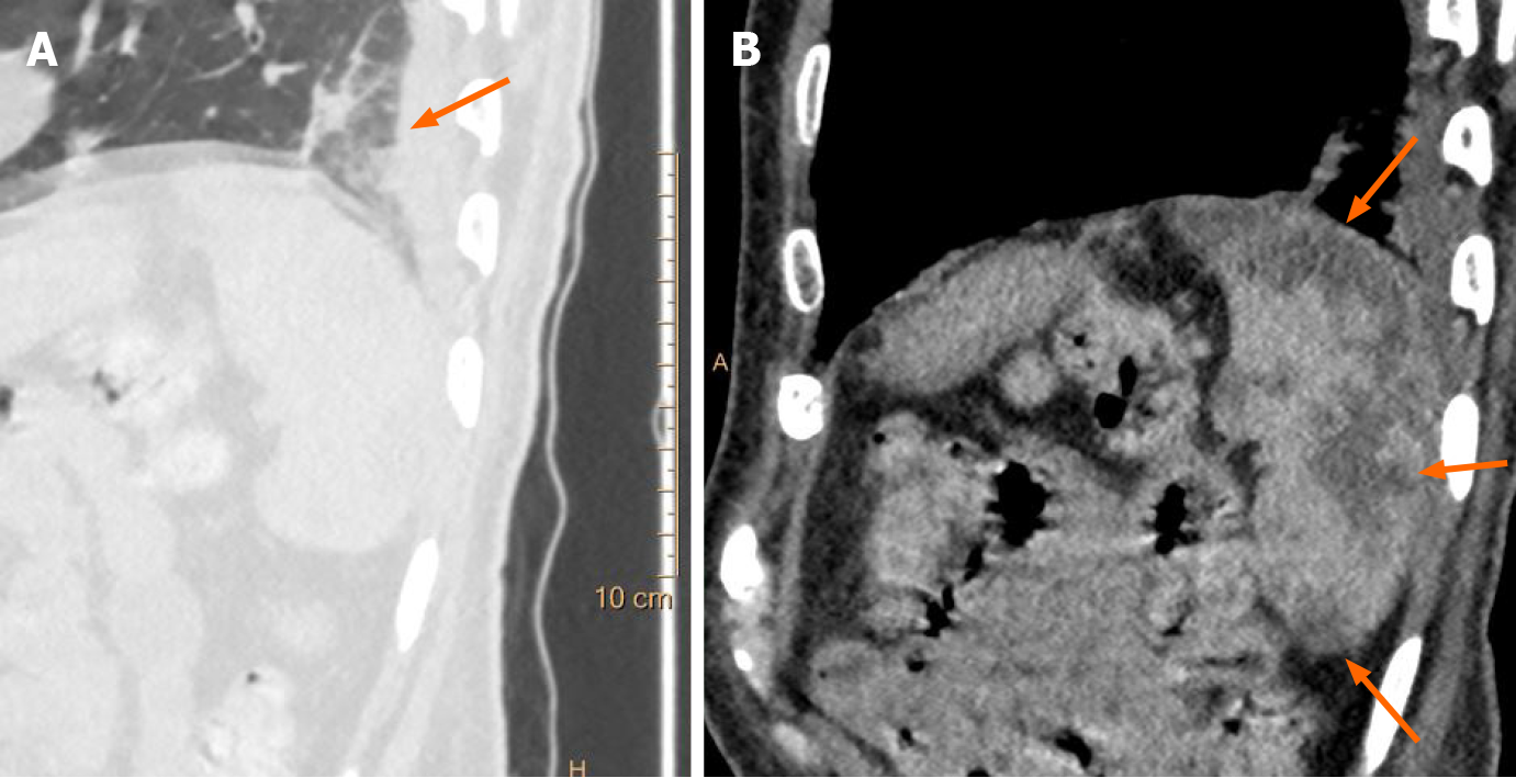
Figure 7 Incidentally detected splenic infarction in a patient with coronavirus disease 2019.
A: Sagittal chest computed tomography (CT) image shows subpleural ground glass opacities in the lung fields in a case of coronavirus disease 2019 (orange arrow); B: Sagittal contrast enhanced CT image of the abdomen in the same patient show incidentally detected, sharply marginated, wedge shaped, hypodense areas in the spleen, suggestive of splenic infarcts (orange arrows).
- Citation: Vaidya T, Nanivadekar A, Patel R. Imaging spectrum of abdominal manifestations of COVID-19. World J Radiol 2021; 13(6): 157-170
- URL: https://www.wjgnet.com/1949-8470/full/v13/i6/157.htm
- DOI: https://dx.doi.org/10.4329/wjr.v13.i6.157