Copyright
©The Author(s) 2021.
World J Radiol. Apr 28, 2021; 13(4): 75-93
Published online Apr 28, 2021. doi: 10.4329/wjr.v13.i4.75
Published online Apr 28, 2021. doi: 10.4329/wjr.v13.i4.75
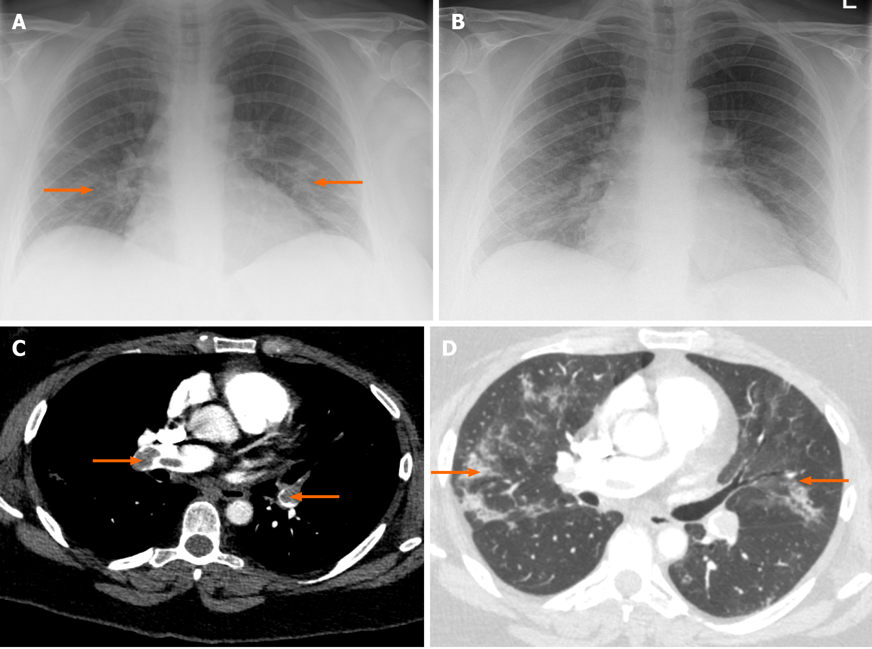
Figure 3 Case 3.
A: The chest radiograph (CXR) at the time of admission showing mild bilateral opacities (arrows); B: CXR on 2nd admission with improving bibasilar lung infiltrates; C: Computed tomographic pulmonary angiogram (CTPA) of case 3 showing evidence of extensive pulmonary embolism (arrows); D: The CTPA lung window showing resolving coronavirus disease-19 pneumonia (arrows).
- Citation: Kumar H, Fernandez CJ, Kolpattil S, Munavvar M, Pappachan JM. Discrepancies in the clinical and radiological profiles of COVID-19: A case-based discussion and review of literature. World J Radiol 2021; 13(4): 75-93
- URL: https://www.wjgnet.com/1949-8470/full/v13/i4/75.htm
- DOI: https://dx.doi.org/10.4329/wjr.v13.i4.75


