
















©The Author(s) 2020.
World J Radiol. Aug 28, 2020; 12(8): 172-183
Published online Aug 28, 2020. doi: 10.4329/wjr.v12.i8.172
Published online Aug 28, 2020. doi: 10.4329/wjr.v12.i8.172
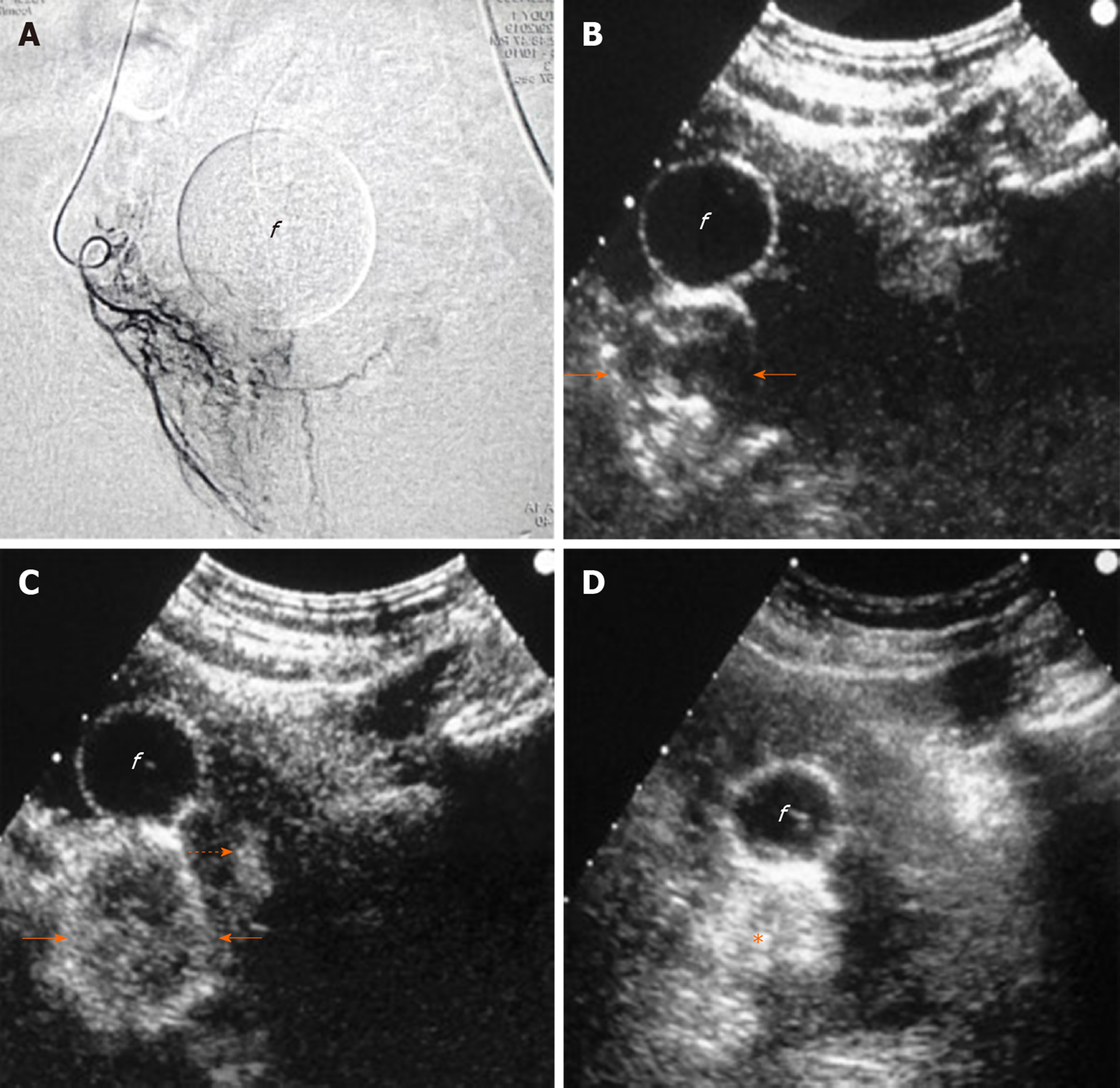
Figure 6 Intraprocedural sonographic evaluation of prostatic artery embolization with intraarterial contrast-enhanced ultrasonography.
A: Angiographic image (anteroposterior projection) shows the right prostatic artery and its branches; B: Transabdominal contrast-enhanced ultrasonography (t.a.-CEUS) image 3 s after intraarterial injection of diluted Sonovue shows rapid, intense enhancement of the right prostatic lobe, more prominent at its periphery (arrows); C: t.a.-CEUS image 7 s later, shows more diffuse enhancement of the right lobe and subtle enhancement in a small area of the left lobe (dotted arrow). No signs of extraprostatic enhancement were noticed and embolization was performed with microspheres (diameter: 100-300 μm); D: Unenhanced t.a.-US image 10 min post-embolization shows increased echogenicity of the right lobe (asterisk). “f” indicates the balloon of the Foley catheter in all images. CEUS images are in axial plane.
- Citation: Moschouris H, Dimakis A, Anagnostopoulou A, Stamatiou K, Malagari K. Sonographic evaluation of prostatic artery embolization: Far beyond size measurements. World J Radiol 2020; 12(8): 172-183
- URL: https://www.wjgnet.com/1949-8470/full/v12/i8/172.htm
- DOI: https://dx.doi.org/10.4329/wjr.v12.i8.172