Copyright
©The Author(s) 2019.
World J Radiol. Sep 28, 2019; 11(9): 116-125
Published online Sep 28, 2019. doi: 10.4329/wjr.v11.i9.116
Published online Sep 28, 2019. doi: 10.4329/wjr.v11.i9.116
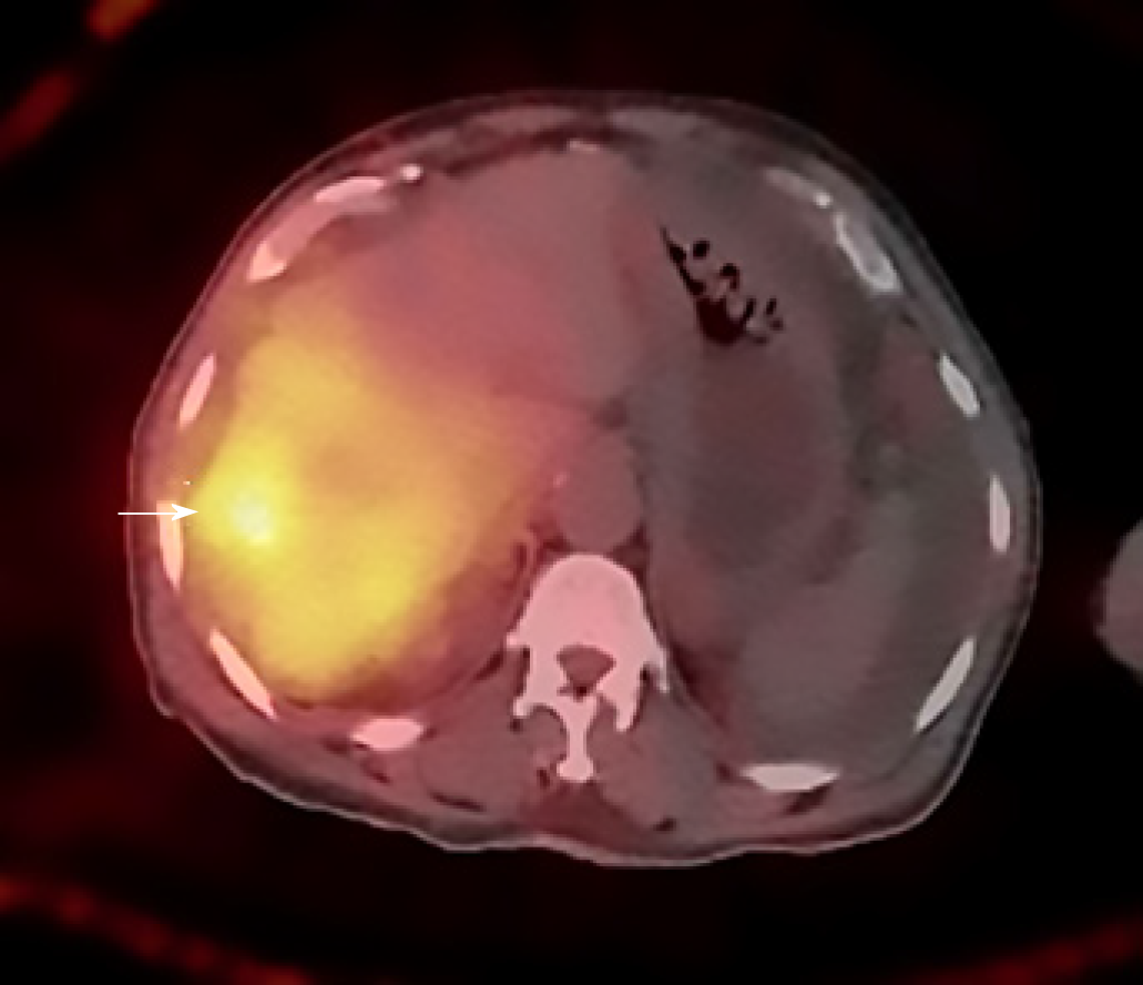
Figure 6 Axial post procedure single photon emission tomography image shows appropriate distribution of Yttrium 90 particles in the right hepatic lobe with increased focal uptake in one of the right lobe lesions (white arrow).
- Citation: Bhat AP, Schuchardt PA, Bhat R, Davis RM, Singh S. Metastatic appendiceal cancer treated with Yttrium 90 radioembolization and systemic chemotherapy: A case report. World J Radiol 2019; 11(9): 116-125
- URL: https://www.wjgnet.com/1949-8470/full/v11/i9/116.htm
- DOI: https://dx.doi.org/10.4329/wjr.v11.i9.116


