
















©The Author(s) 2019.
World J Radiol. Aug 28, 2019; 11(8): 110-115
Published online Aug 28, 2019. doi: 10.4329/wjr.v11.i8.110
Published online Aug 28, 2019. doi: 10.4329/wjr.v11.i8.110
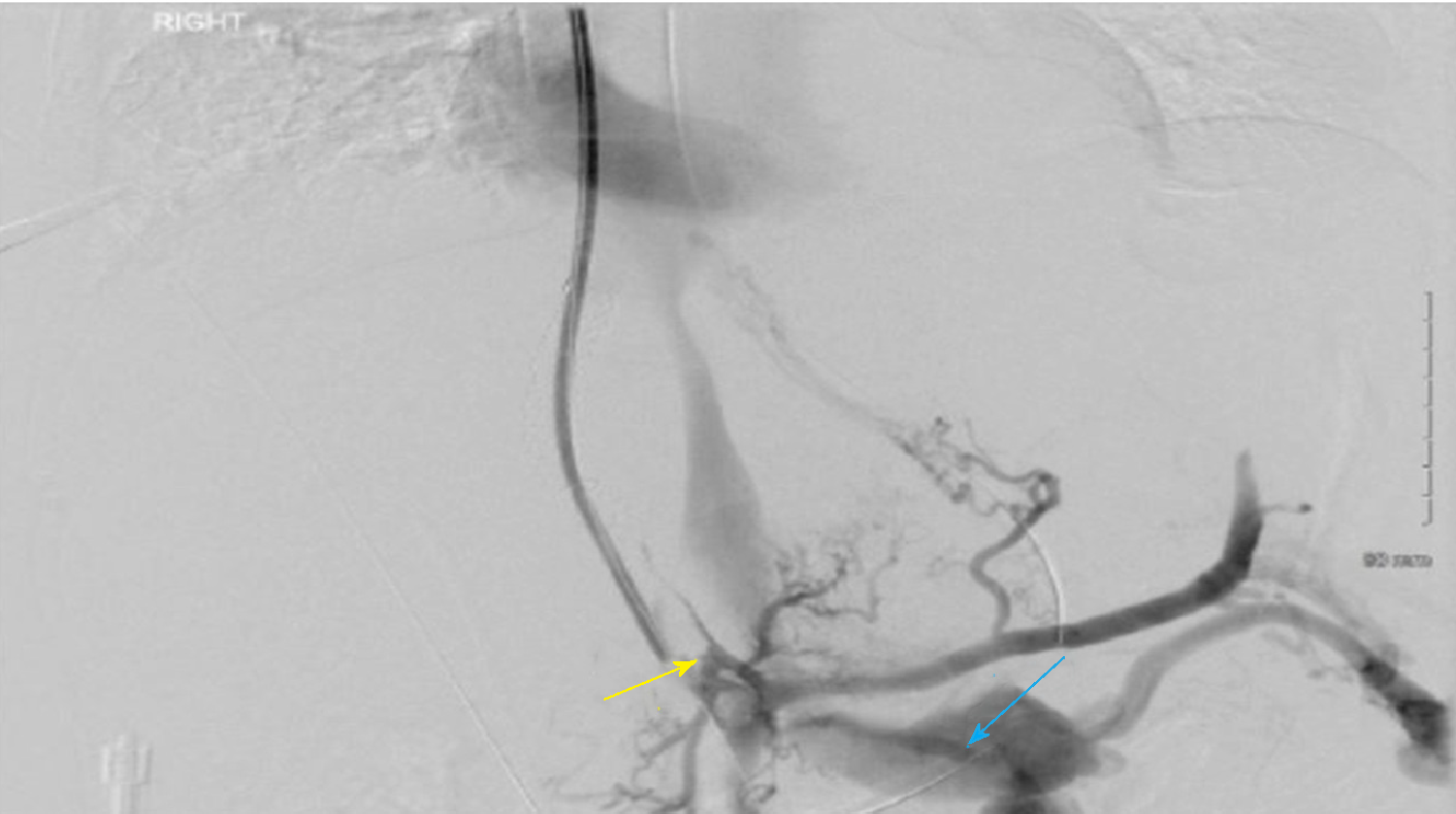
Figure 3 Angiographic image showing filling defect in the transjugular intrahepatic portosystemic shunt and the portal vein (yellow arrow) with portosystemic shunting to left renal vein (blue arrow).
- Citation: Anand R, Ali SE, Raissi D, Frandah WM. Duodenal variceal bleeding with large SPSS treated with transjugular intrahepatic portosystemic shunt and embolization: A case report. World J Radiol 2019; 11(8): 110-115
- URL: https://www.wjgnet.com/1949-8470/full/v11/i8/110.htm
- DOI: https://dx.doi.org/10.4329/wjr.v11.i8.110