Copyright
©The Author(s) 2019.
World J Radiol. Jun 28, 2019; 11(6): 81-93
Published online Jun 28, 2019. doi: 10.4329/wjr.v11.i6.81
Published online Jun 28, 2019. doi: 10.4329/wjr.v11.i6.81
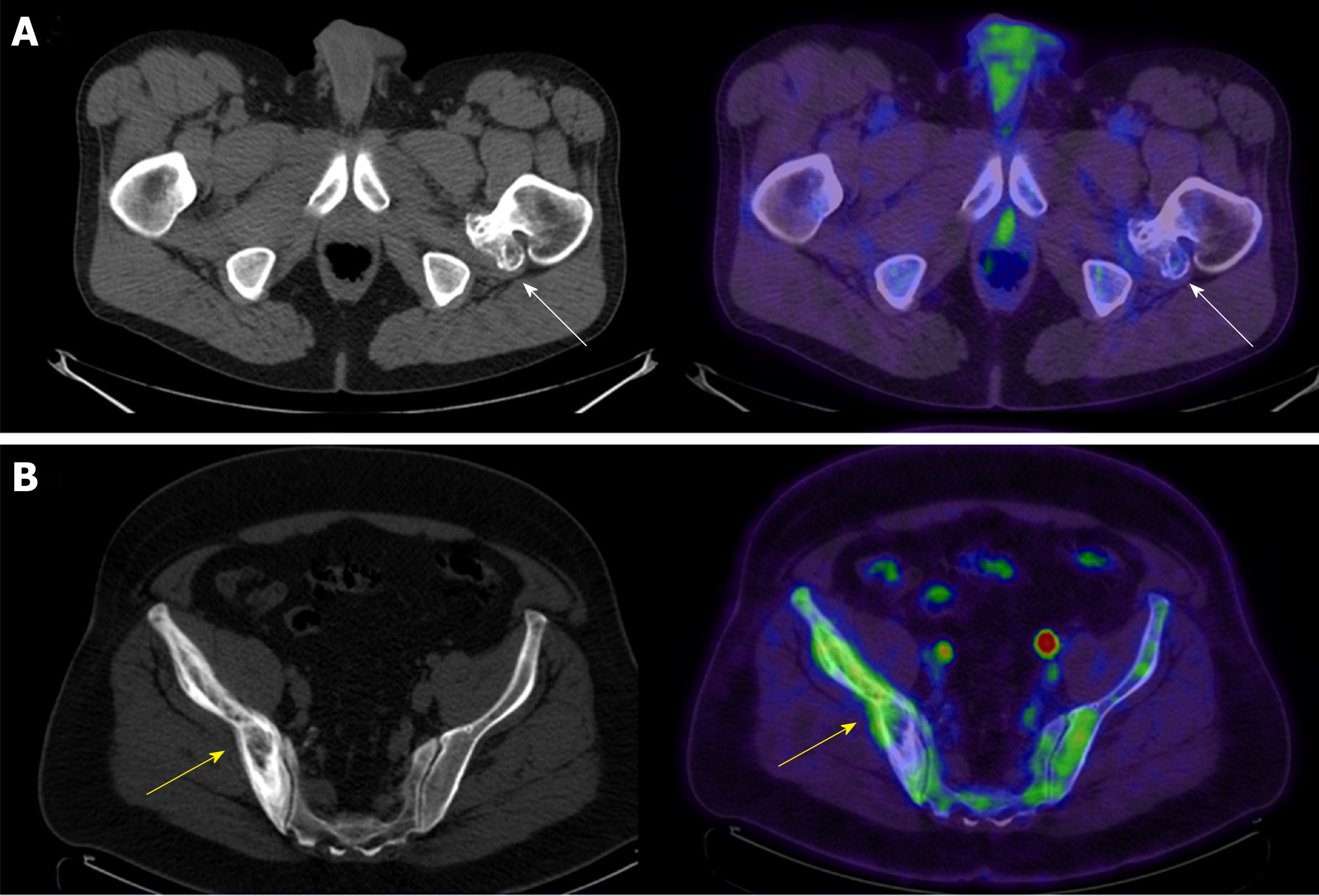
Figure 5 Axial computed tomography and axial fused positron emission tomography/computed tomography of two patients.
A: Axial computed tomography (CT) and axial fused positron emission tomography (PET)/CT of the pelvis in a 51-year-old male with diffuse large B-cell lymphoma and a sessile osteochondroma arising from the proximal left femur (arrows) [maximum standardized uptake value (SUV) 1.64]; B: Axial CT and axial fused PET/CT of the pelvis in a 61-year-old male with a history of a follicular lymphoma and Paget’s disease of the bone involving the right ilium (arrows) (maximum SUV 3.92).
- Citation: Elangovan SM, Sebro R. Positron emission tomography/computed tomography imaging appearance of benign and classic “do not touch” osseous lesions. World J Radiol 2019; 11(6): 81-93
- URL: https://www.wjgnet.com/1949-8470/full/v11/i6/81.htm
- DOI: https://dx.doi.org/10.4329/wjr.v11.i6.81


