Copyright
©The Author(s) 2018.
World J Radiol. Jul 28, 2018; 10(7): 65-77
Published online Jul 28, 2018. doi: 10.4329/wjr.v10.i7.65
Published online Jul 28, 2018. doi: 10.4329/wjr.v10.i7.65
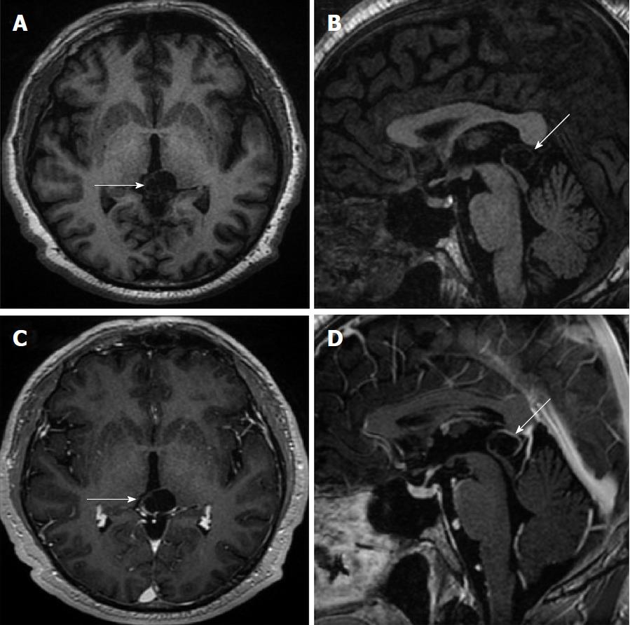
Figure 5 Fifty years old male patient with atypical pineal cyst (patient No.
40). Axial plane (A) sagittal plane T1 weighted (B) axial plane (C) sagittal plane contrast-enhanced magnetic resonance imaging (D). A, B, C and D: Bilocular, lobule contoured atypical pineal cyst is shown in pineal area (white arrows); C and D: Partial rim-like enhancement is shown (white arrows).
- Citation: Gokce E, Beyhan M. Evaluation of pineal cysts with magnetic resonance imaging. World J Radiol 2018; 10(7): 65-77
- URL: https://www.wjgnet.com/1949-8470/full/v10/i7/65.htm
- DOI: https://dx.doi.org/10.4329/wjr.v10.i7.65


