Copyright
©The Author(s) 2018.
World J Radiol. Apr 28, 2018; 10(4): 30-45
Published online Apr 28, 2018. doi: 10.4329/wjr.v10.i4.30
Published online Apr 28, 2018. doi: 10.4329/wjr.v10.i4.30
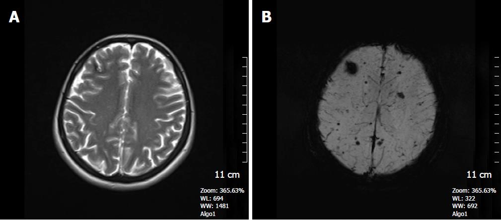
Figure 5 A 41-year-old woman who had a history of familial cavernous malformation underwent magnetic resonance imaging screening.
A: Axial FSE T2W image, a right frontal subcortical small cavernous malformation is seen on this image; B: SWI minIP image, numerous tiny cavernous malformations throughout the brain parenchyma is detected. FSE T2W image is unable to show these lesions. The patient was considered to have familial cerebral cavernous malformation. SWI: Susceptibility weighted imaging; FSE: Fast spin echo.
- Citation: Halefoglu AM, Yousem DM. Susceptibility weighted imaging: Clinical applications and future directions. World J Radiol 2018; 10(4): 30-45
- URL: https://www.wjgnet.com/1949-8470/full/v10/i4/30.htm
- DOI: https://dx.doi.org/10.4329/wjr.v10.i4.30


