Copyright
©The Author(s) 2018.
World J Radiol. Apr 28, 2018; 10(4): 30-45
Published online Apr 28, 2018. doi: 10.4329/wjr.v10.i4.30
Published online Apr 28, 2018. doi: 10.4329/wjr.v10.i4.30
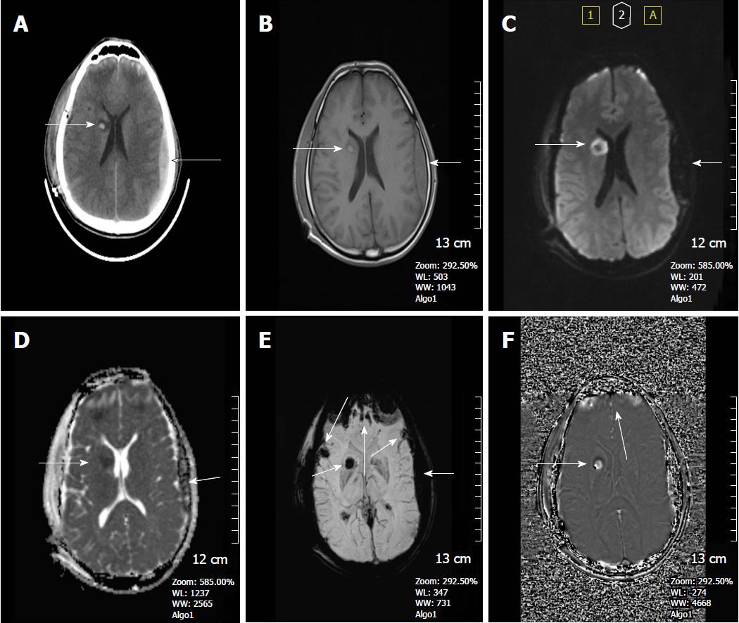
Figure 3 A 37-year-old man who had an accident was in coma after traumatic brain injury.
A: On the non-contrast CT image, bilateral frontal subcortical and right basal ganglia hyperdense hemorrhagic foci with surrounding hypodense edema are seen consistent with diffuse axonal injury. Also left parieto-temporal subdural hemorrhage is present. Post-op changes are present on the right with a tiny frontal subdural hematoma; B: SE T1W image, can only reveal hyperintense right basal ganglia hemorrhagic lesion with surrounding hypointense edema and left subdural hemorrhage, but can not demonstrate the other parenchymal lesions; C: Diffusion weighted image (DWI) reveals hyperintense caudate lesions; D: Apparent diffusion coefficients (ADC) map demonstrates restricted diffusion within the lesions; E: SWI minIP image, clearly depicts multiple frontal cortical and subcortical and also right basal ganglion microhemorrhages better than those of CT and T1W MR image. The bilateral subdural hematomas are nearly as dark as the cortical bone; F: Phase contrast SWI image, hemorrhagic lesions show a bright/positive shift effect on phase image, due to paramagnetic susceptibility effect. SWI: Susceptibility weighted imaging; minIP: Minimum intensity projection algorithm; SE: Spin echo.
- Citation: Halefoglu AM, Yousem DM. Susceptibility weighted imaging: Clinical applications and future directions. World J Radiol 2018; 10(4): 30-45
- URL: https://www.wjgnet.com/1949-8470/full/v10/i4/30.htm
- DOI: https://dx.doi.org/10.4329/wjr.v10.i4.30


