Copyright
©The Author(s) 2018.
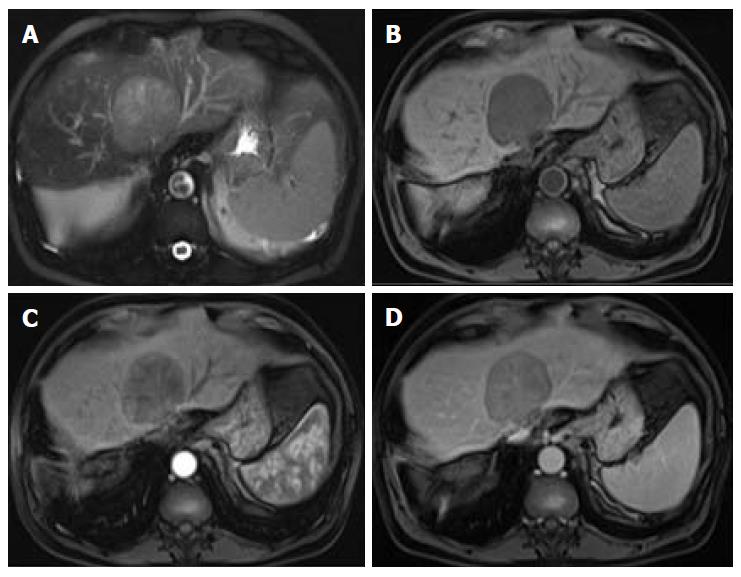
Figure 13 Mosaic architecture.
Fat-suppressed T2-weighted image (A), pre (B) and post-contrast fat-suppressed 3D-GRE T1-weighted images during the late hepatic arterial (C) and delayed (D) phases. A large mass showing randomly distributed internal compartments with different signal intensities on the T2 weighed image (A) and variable enhancement patterns on the late arterial (C) and delayed phases (D). This is an extremely unusual feature in non- hepatocellular carcinoma (HCC) tumors. In large, arterial hyperenhancement might be more difficult to recognize and as in this case and the presence of mosaic architecture aids in the diagnosis of HCC.
- Citation: Campos-Correia D, Cruz J, Matos AP, Figueiredo F, Ramalho M. Magnetic resonance imaging ancillary features used in Liver Imaging Reporting and Data System: An illustrative review. World J Radiol 2018; 10(2): 9-23
- URL: https://www.wjgnet.com/1949-8470/full/v10/i2/9.htm
- DOI: https://dx.doi.org/10.4329/wjr.v10.i2.9


