Copyright
©The Author(s) 2018.
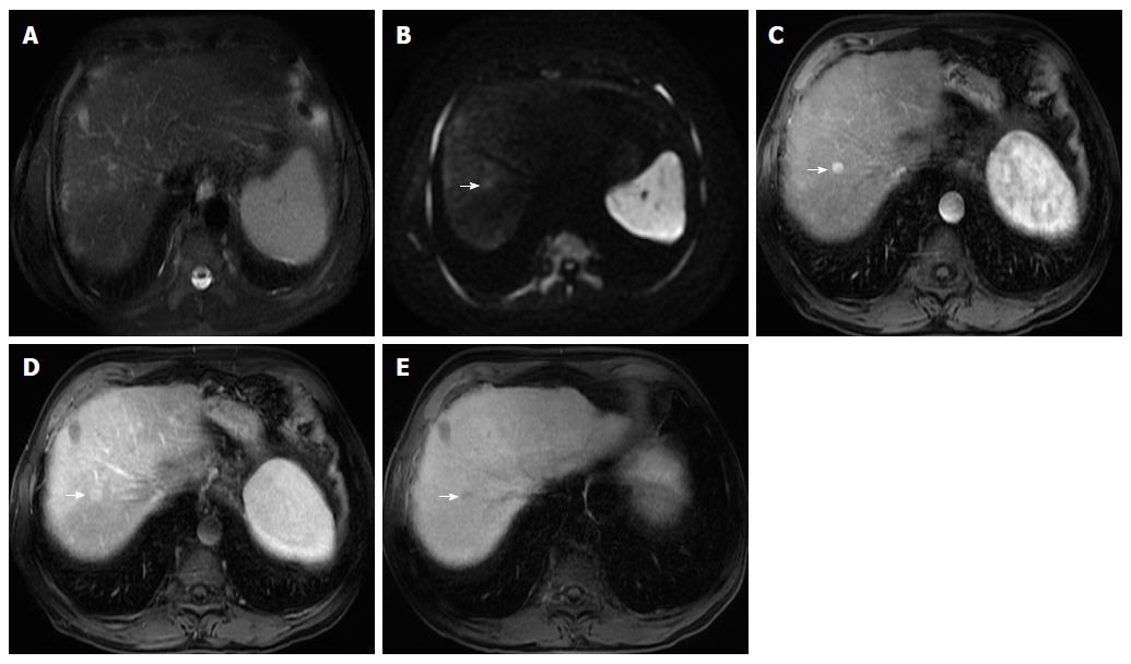
Figure 4 Small hepatocellular carcinoma.
Fat-suppressed T2-weighted image (A), diffusion-weighted image (DWI; B = 600) (B), and hepatobiliary contrast enhanced (gadobenate dimeglumine) fat-suppressed 3D-GRE T1-weighted images in the late hepatic arterial (C), delayed (D), and hepatobiliary phases (E). A small nodule in the right hepatic lobe is seen, showing near isointense signal on T2- weighted image (A) and mild high signal intensity on DWI (arrow, B). An increased arterial enhancement is seen (arrow, C), fading out in the delayed phase (D) and decreased uptake on the hepatobiliary phase (arrow, E). These findings are suspicious for malignancy. This nodule was ablated and histologically proven HCC.
- Citation: Campos-Correia D, Cruz J, Matos AP, Figueiredo F, Ramalho M. Magnetic resonance imaging ancillary features used in Liver Imaging Reporting and Data System: An illustrative review. World J Radiol 2018; 10(2): 9-23
- URL: https://www.wjgnet.com/1949-8470/full/v10/i2/9.htm
- DOI: https://dx.doi.org/10.4329/wjr.v10.i2.9


