Copyright
©2009 Baishideng Publishing Group Co.
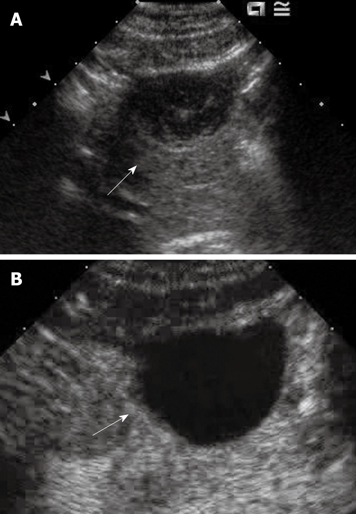
Figure 7 Complicated cyst.
A: A slightly hypo-echoic lesion (arrow); B: CEUS at the portal phase of the lesion shows lack of contrast enhancement (arrow), as well as throughout the remaining vascular phases (not shown). Some internal non-enhancing debris within the cyst is still appreciable.
- Citation: Wong GLH, Xu HX, Xie XY. Detection Of Focal Liver Lesions In Cirrhotic Liver Using Contrast-Enhanced Ultrasound. World J Radiol 2009; 1(1): 25-36
- URL: https://www.wjgnet.com/1949-8470/full/v1/i1/25.htm
- DOI: https://dx.doi.org/10.4329/wjr.v1.i1.25


