Copyright
©The Author(s) 2017.
World J Radiol. Dec 28, 2017; 9(12): 426-437
Published online Dec 28, 2017. doi: 10.4329/wjr.v9.i12.426
Published online Dec 28, 2017. doi: 10.4329/wjr.v9.i12.426
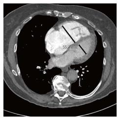
Figure 1 Right/left ventricular ratio greater than 0.
9, measured in the basal portion of the ventricles at the level of the atrioventricular valve.
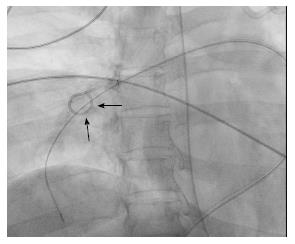
Figure 2 A pigtail catheter in the right pulmonary artery is rotated to fragment and then disperse clot into the larger volume peripheral circulation.
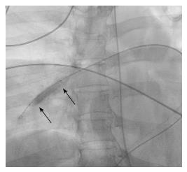
Figure 3 An angioplasty balloon catheter in the right pulmonary artery being used to fragment and disperse clot into the larger volume peripheral circulation.
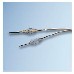
Figure 4 Trerotola thrombolytic device (Permission to use image granted by Teleflex Inc.
, Wayne, PA, United States).
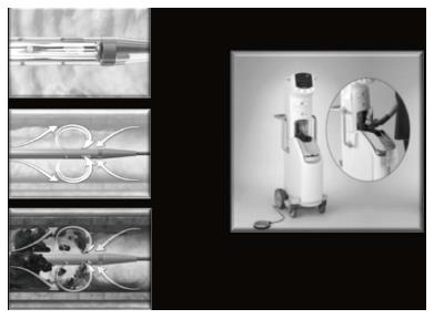
Figure 5 Angiojet rheolytic thrombectomy device (Boston Scientific, Marlborough, MA, United States).
Distal Saline jets fragment clots and high velocity saline loops into a second lumen, aspirating fragments of clot into the sideports through the Venturi effect (Image provided courtesy of Boston Scientific. ©2017 Boston Scientific Corporation or its affiliates. All rights reserved.
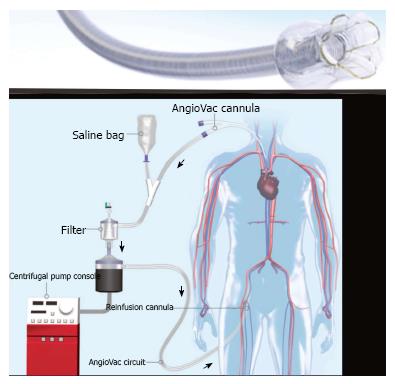
Figure 6 Angiovac catheter (Angiodynamics, Inc.
, Latham, NY, United States) and reperfusion circuit, in which aspirated clot is filtered from aspirated blood, which is subsequently returned to the patient (permission to use image granted by Angiodynamics, Inc.).
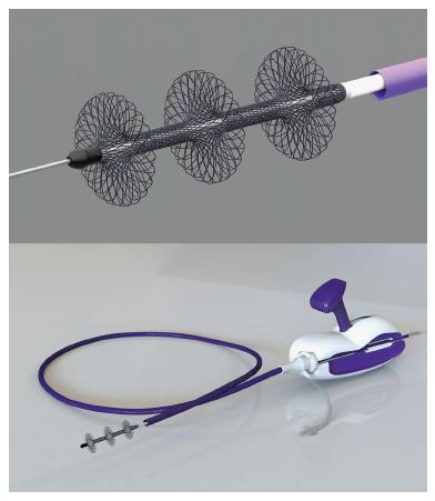
Figure 7 Flowtriever device (Inari Medical, Irvine, CA, United States) with three braided nitinol disks to engage clot and pull it into the aspiration guide catheter for evacuation by the retraction aspirator (Permission to use image granted by Inari Medical).
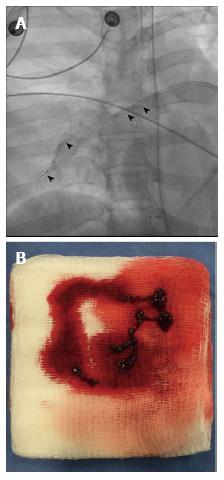
Figure 8 Penumbra device.
A: Penumbra Indigo CAT-8 (Penumbra, Inc., Alameda, CA, United States) in the right pulmonary artery. The guiding catheter (arrowheads) is in the right main pulmonary artery and the aspiration catheter (arrows) is in the right lower lobe pulmonary artery. The separator wire used to promote aspiration is exiting the aspiration catheter; B: Small volume clot aspirated with the Penumbra Indigo CAT-8 system.
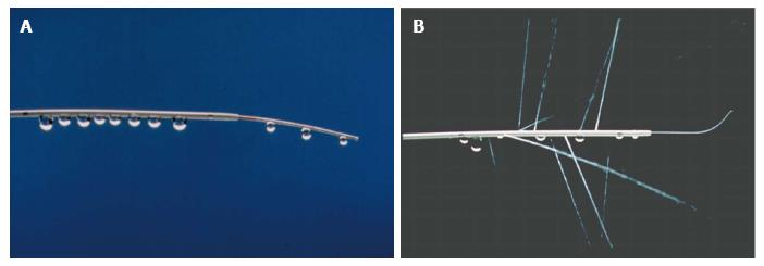
Figure 9 Multiholed infusion catheter.
A: A multiholed infusion catheter with sideholes dispersing infusate within clot and infusion wire further distributing the infusate while also occluding the endhole of the infusion catheter, forcing flow out of the sideholes of the catheter (Permission to use image granted by Angiodynamics, Inc.); B: A multiholed infusion catheter with sideholes dispersing infusate within clot and infusion wire further distributing the infusate while also occluding the endhole of the infusion catheter, forcing flow out the sideholes of the catheter (Permission to use image granted by Angiodynamics, Inc.).
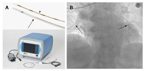
Figure 10 Ekos system.
A: Ekos system with multiholed infusion catheter (arrow), ultrasonic core transducer (arrowhead) and control unit (Permission to use image granted by Ekos Corp., Bothel, WA, United States); B: Ekos catheters (arrows) with ultrasonic core transducers in place in the right and left pulmonary arteries.
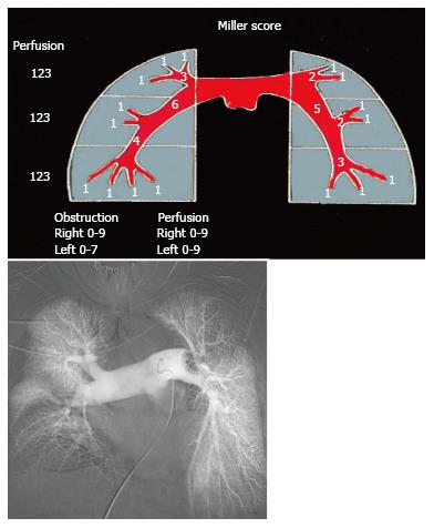
Figure 11 Miller Score.
Pulmonary arteriogram demonstrating left main pulmonary artery embolus (obstruction score 7) and severely reduced perfusion of the upper and middle zones of the left lung (perfusion index 2 + 2). There is occlusion of the left lower lobe pulmonary and middle lobe pulmonary artery (obstructive score 4 + 2) with perfusion severely reduced in the lower lobe and mildly reduced in the middle lobe (perfusion index 2 + 1). This patients combined Miller score is therefore 20.
- Citation: Nosher JL, Patel A, Jagpal S, Gribbin C, Gendel V. Endovascular treatment of pulmonary embolism: Selective review of available techniques. World J Radiol 2017; 9(12): 426-437
- URL: https://www.wjgnet.com/1949-8470/full/v9/i12/426.htm
- DOI: https://dx.doi.org/10.4329/wjr.v9.i12.426


