Copyright
©The Author(s) 2015.
World J Radiol. Jul 28, 2015; 7(7): 149-156
Published online Jul 28, 2015. doi: 10.4329/wjr.v7.i7.149
Published online Jul 28, 2015. doi: 10.4329/wjr.v7.i7.149
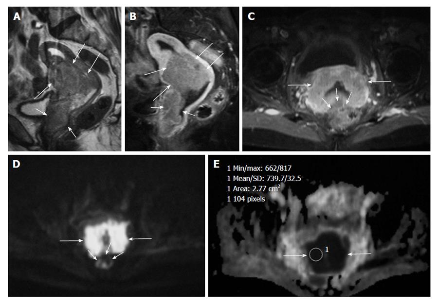
Figure 1 Forty-three years old woman with stage III squamous cell carcinoma of the uterine cervix invading vagina.
A: Sagittal T2-weighted image of the uterus shows cervical cancer (long arrows) extending both to the corpus uteri and vagina (short arrows); B: Sagittal contrast-enhanced T1-weighted image with fat suppression shows enhancing cervical cancer (arrows). The tumor invades anterior vaginal wall (short arrows); C: Axial contrast-enhanced T1-weighted image with fat suppression shows enhancing cervical cancer (long arrows). There is suspicious invasion of the mass to the rectum (short arrows); D: Diffusion-weighted imaging with b = 1000 s/mm2 clearly shows a well-defined hyperintensity mass in the cervical area with no invasion to rectum (short arrows); E: On the apparent diffusion coefficient (ADC) map the tumor is hypointense (arrows). The ADC value within the mass is 0.73 × 10⁻³ mm2/s.
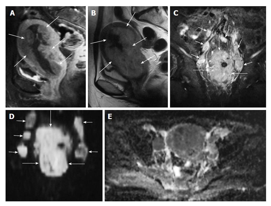
Figure 2 A 57-year-old woman with endometrial carcinoma.
A: Sagittal contrast-enhanced T1-weighted image with fat suppression shows enhancing endometrial cancer with infiltration of myometrium (arrows); B: Sagittal T2-weighted image demonstrating hyperintense endometrial cancer with infiltration of myometrium (arrows); C: Coronal fat supressed T2-weighted image reveals a tumor in the corpus uteri (long arrows), and bilateral metastatic lymphadenopathies along the iliac chains (short arrows); D: Coronal DWI (b = 1000 s/mm2) shows a marked hyperintense tumor in the corpus uteri (long arrows), and bilateral metastatic lymphadenopathies along the iliac chains (short arrows); E: Axial apparent diffusion coefficient map reveals the right sided metastatic lymph node and endometrium with restricted diffusion.
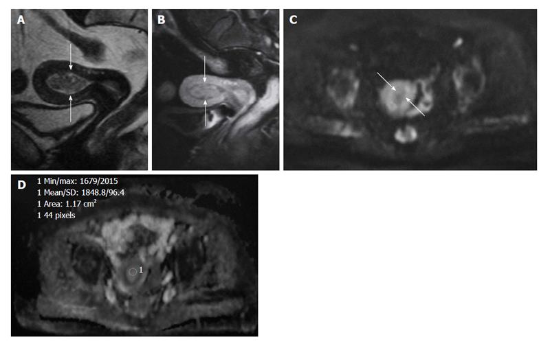
Figure 3 A 42-year-old woman with endometrial polyp.
A: Hypointense polyp in the endometrial cavity on sagittal T2-weighted image mimicking low grade endometrial carcinoma (arrows); B: Sagittal contrast-enhanced T1-weighted image with fat suppression shows enhancing endometrial polyp (arrows); C: On the axial DWI (b = 1000 s/mm2) image, the mass is hypointense clearly excluding malignancy (arrows); D: Corresponding axial apparent diffusion coefficient (ADC) map. The ADC value within the mass is 1.85 × 10⁻³ mm2/s.
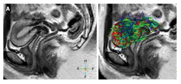
Figure 4 Thirty two years old volunteer.
A: Sagittal T2-weighted image of a normal uterus; B: 3D whole tractography image of the normal uterus. Red colors represent a right-left orientation, blue represents a cranio-caudal orientation and green represents an antero-posterior orientation of diffusion. Changes in the intensity of the color represent different strengths of anisotropy.
- Citation: Kara Bozkurt D, Bozkurt M, Nazli MA, Mutlu IN, Kilickesmez O. Diffusion-weighted and diffusion-tensor imaging of normal and diseased uterus. World J Radiol 2015; 7(7): 149-156
- URL: https://www.wjgnet.com/1949-8470/full/v7/i7/149.htm
- DOI: https://dx.doi.org/10.4329/wjr.v7.i7.149


