Copyright
©The Author(s) 2024.
World J Radiol. Mar 28, 2024; 16(3): 58-68
Published online Mar 28, 2024. doi: 10.4329/wjr.v16.i3.58
Published online Mar 28, 2024. doi: 10.4329/wjr.v16.i3.58
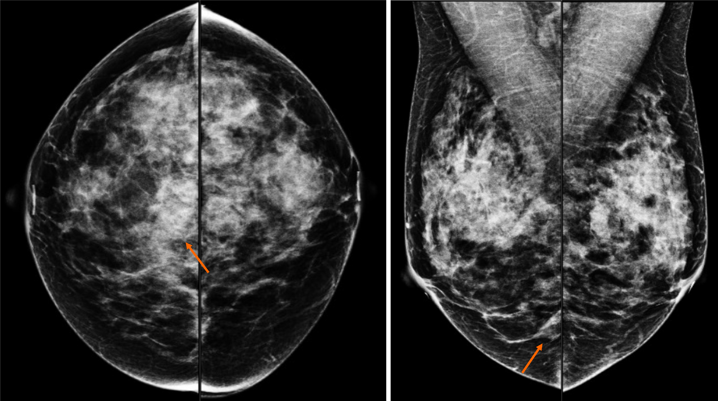
Figure 1 Craneocaudal and Mediolateral Oblique projections of mammography.
The breast tissue is extremely dense (category “D” of the American College of Radiology, 2013). In the right breast exists a conglomerate of nodules (arrow) localized at the third posterior of the union of lower quadrants, they are two isodense and oval nodules with obscured margins.
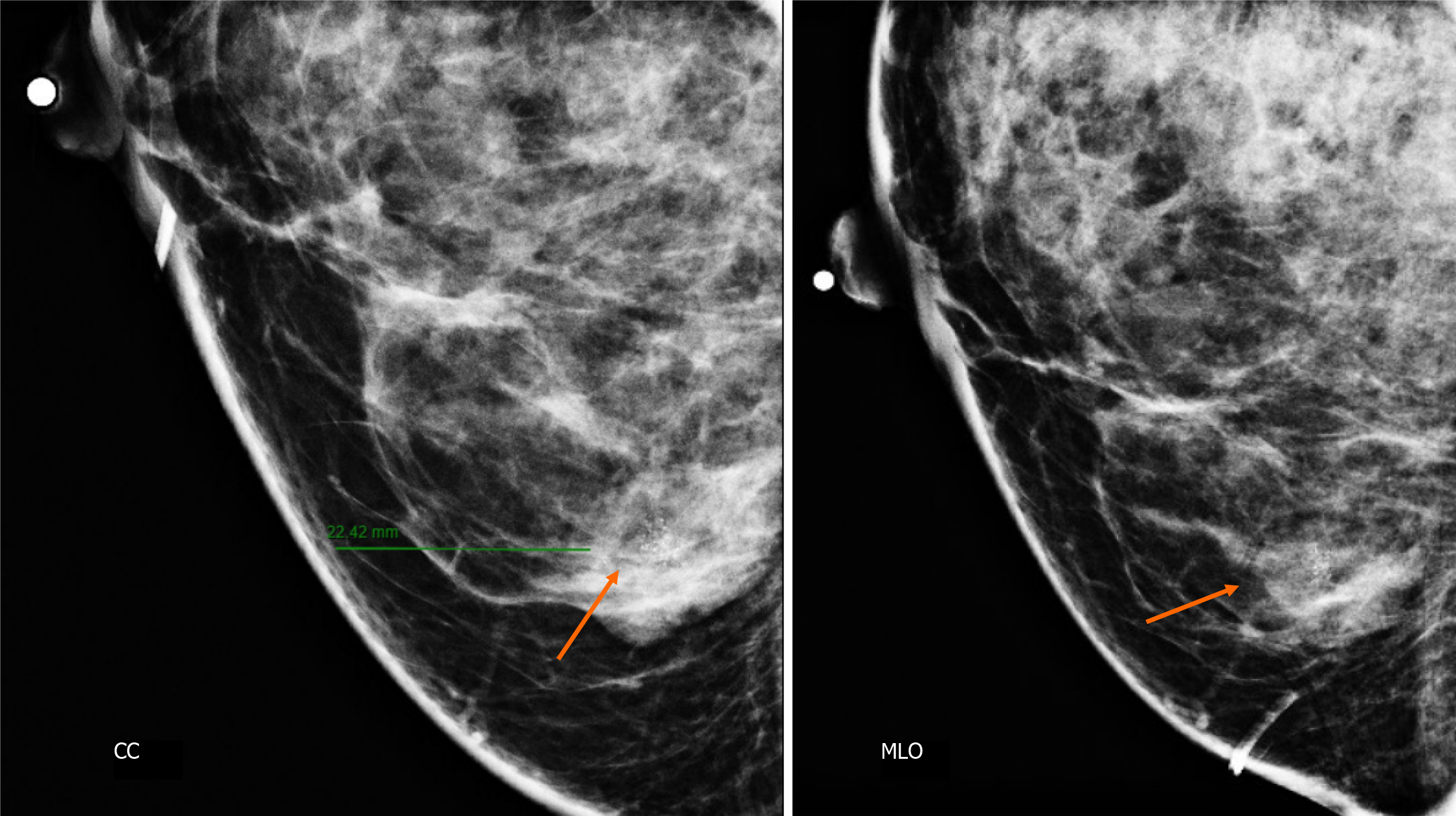
Figure 2 Craniocaudal and mediolateral oblique projections with magnification of right breast.
The two confluence nodules are associated with suspicious microcalcifications (arrow). The findings are localized 2 cm from skin and are adjacent to mastoplasty surgical scar indicated by the linear tissue marker as it is seen in the mediolateral oblique projection. CC: Craniocaudal; MLO: Mediolateral oblique.
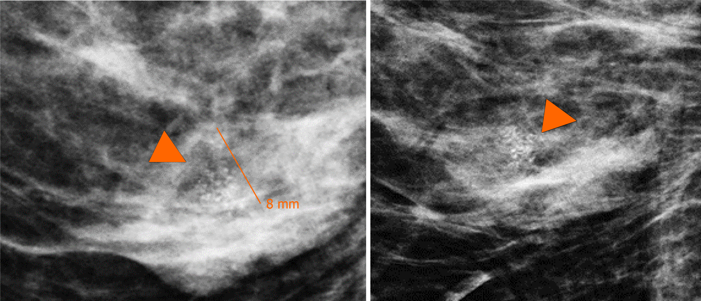
Figure 3 Magnification views in orthogonal projections.
One of the nodules is associated with fine and linear pleomorphic microcalcifications (head arrow) that extended in an area of 8 mm.
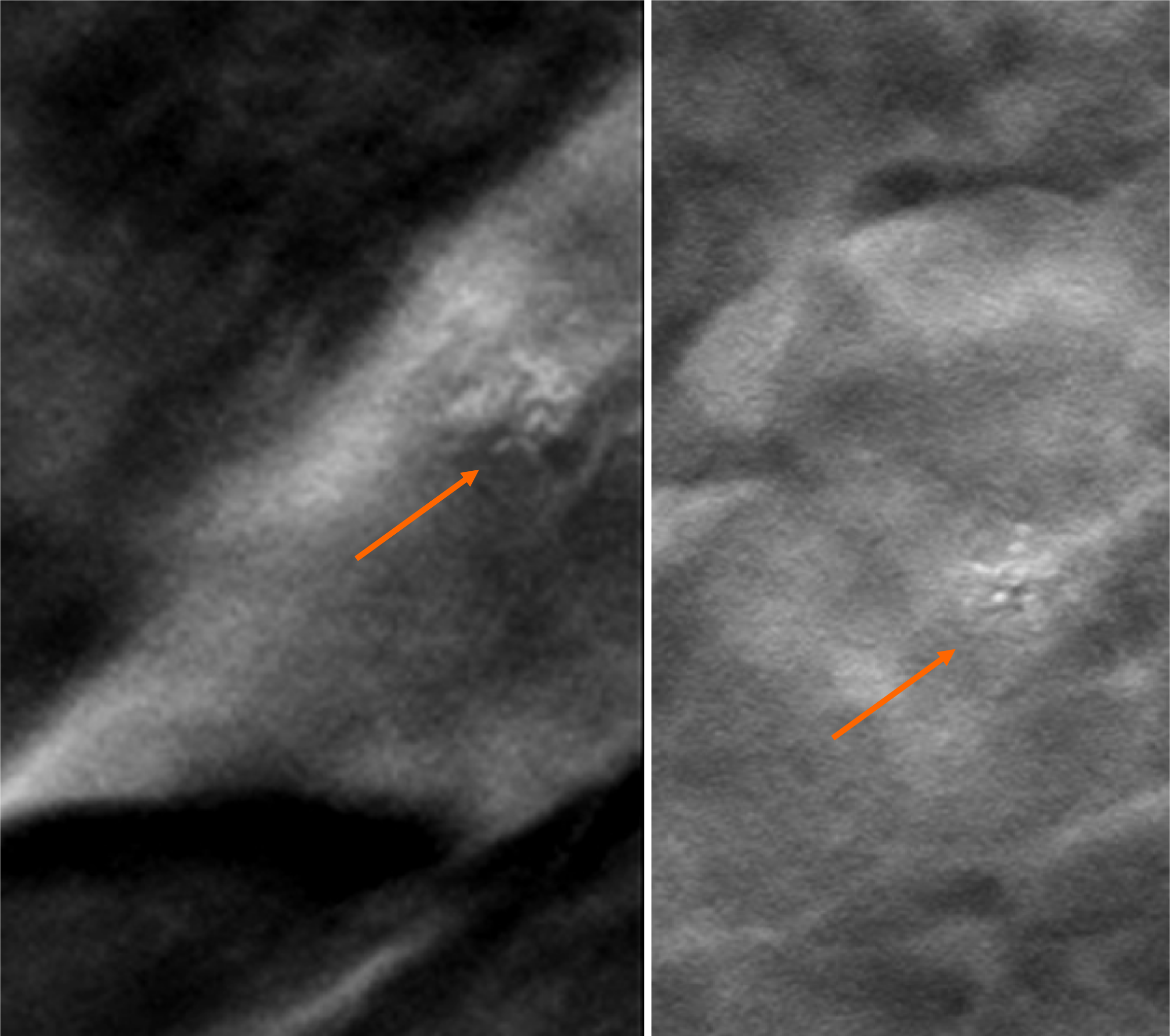
Figure 4 Digital breast tomosynthesis with magnification of craniocaudal and mediolateral oblique projections.
The morphology of microcalcifications is corroborated as fine and linear pleomorphic appearance (arrow).
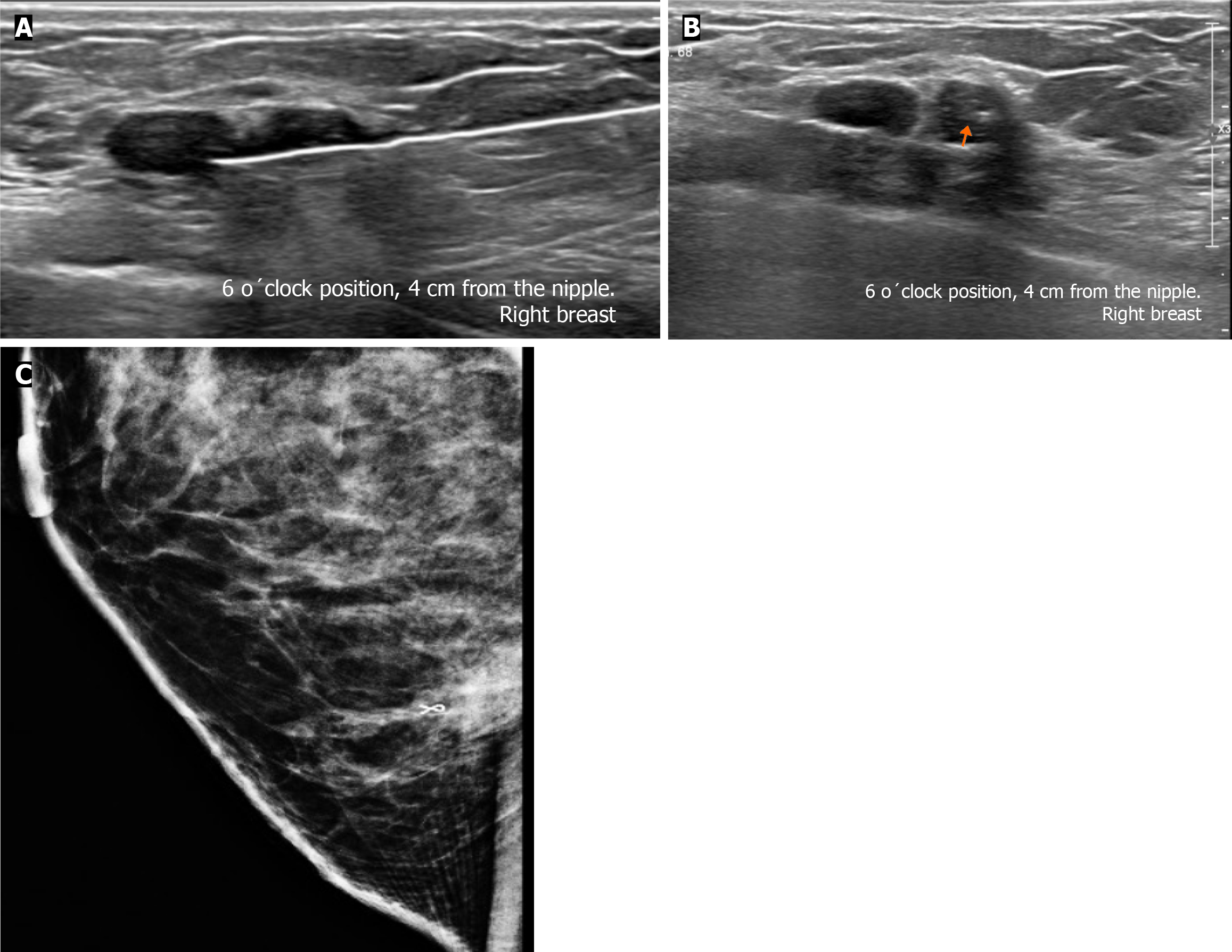
Figure 5 Gray-scale ultrasound and Doppler color images.
A: In the right breast is seen a conglomerate of two solid nodules that are hypoechoic, oval, circumscribed and non-palpable with lateral acoustic shadowing, located at the 6 o'clock position, 4 cm from the nipple in the right breast. In one of the nodules, internal hyperechoic foci are seen, corresponding to microcalcifications shown in mammography; B: The nodules have neither internal nor peripherical vascularity.
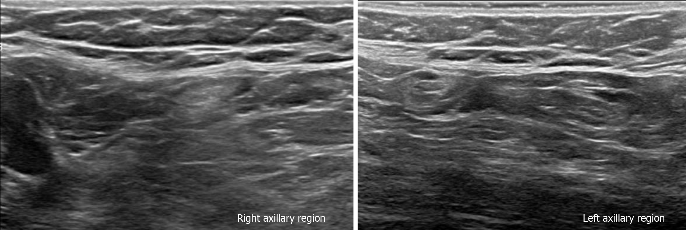
Figure 6 Gray-scale ultrasound images.
The bilateral axillary region shows lymph nodes with conservative morphology and fat hilum, with cortex of < 3 mm.
Figure 7 Ultrasound-guided core biopsy images.
A: Percutaneous biopsy was performed of the conglomerate solid nodules, using a 12-gauze needle; B: A tissue marker or clip is placed in one of the nodules (arrow) which is associated with microcalcifications; C: Lateral projection of mammography. The tissue marker confirms the position of biopsied microcalcifications.
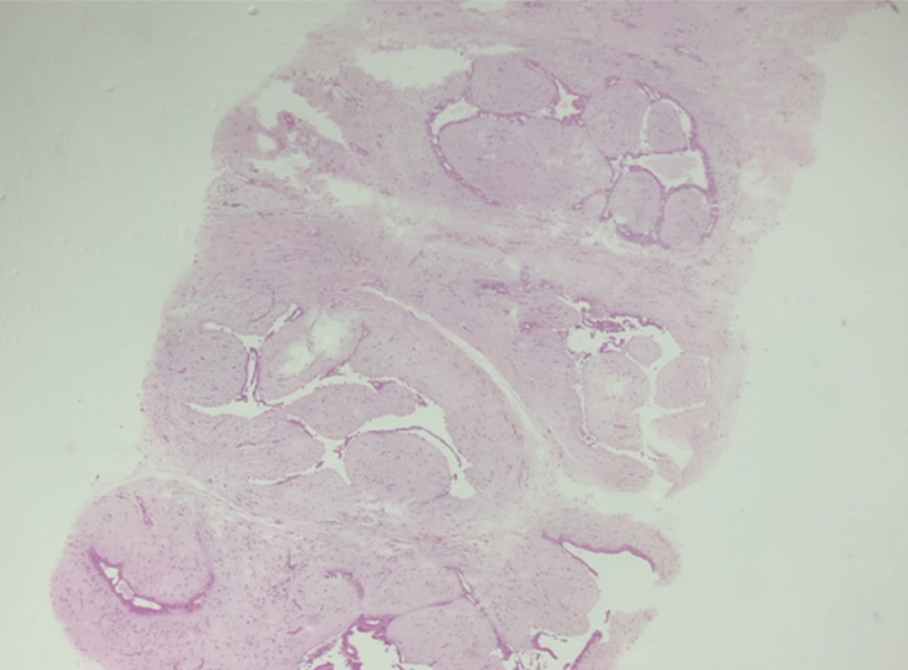
Figure 8 Hematoxylin and eosin stain slide.
Pleomorphic neoplastic cells are seen within a fibroadenoma.
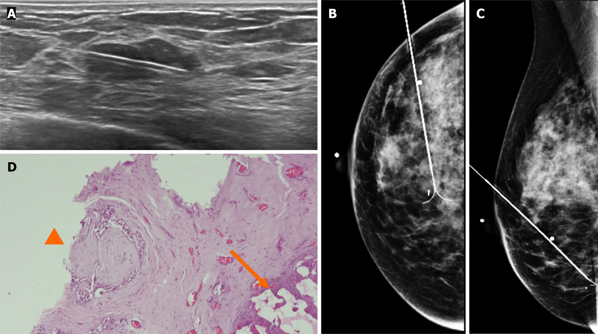
Figure 9 Gray-scale ultrasound images.
A: Preoperative guidewire is inserted percutaneously into the breast to localize the non-palpable conglomerate of nodules done under ultrasound; B and C: Craniocaudal and mediolateral oblique projection. The adequate position of the hook wire is confirmed in the target lesion; D: Histological slide of partial mastectomy. The site of the previous biopsy is seen (arrow). Also, fibroadenoma is shown which contains pleomorphic neoplastic cells and nucleolus with pleomorphic appearance (head arrow) that corresponds to ductal carcinoma in situ.
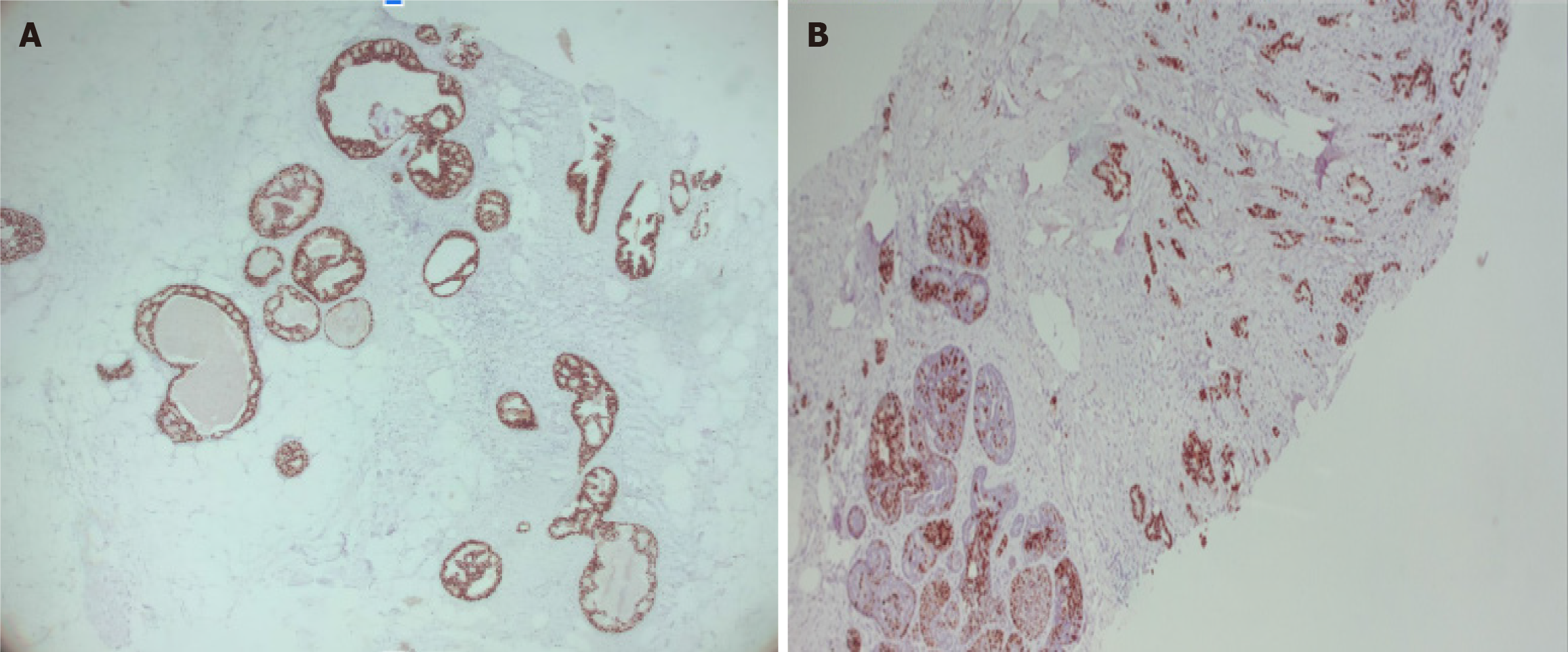
Figure 10 Immunohistochemical staining.
A: Immunohistochemical markers 5 ×, estrogen receptor + (90%) in ductal carcinoma in situ within fibroadenoma; B: Immunohistochemical markers 5 ×, progesterone receptor (+) 70% in ductal carcinoma in situ within fibroadenoma.
- Citation: Olivares-Antúnez Y, Dávila-Zablah YJ, Vázquez-Ávila JR, Gómez-Macías GS, Mireles-Aguilar MT, Garza-Montemayor ML. Ductal carcinoma in situ within a fibroadenoma: A case report and review of literature. World J Radiol 2024; 16(3): 58-68
- URL: https://www.wjgnet.com/1949-8470/full/v16/i3/58.htm
- DOI: https://dx.doi.org/10.4329/wjr.v16.i3.58


