Published online Apr 26, 2016. doi: 10.4330/wjc.v8.i4.323
Peer-review started: October 8, 2015
First decision: December 4, 2015
Revised: December 22, 2015
Accepted: January 5, 2016
Article in press: January 7, 2016
Published online: April 26, 2016
Processing time: 193 Days and 13.7 Hours
Persistent left superior vena cava (LSVC) is a congenital anomaly with 0.3%-1% prevalence in the general population. It is usually asymptomatic but in case of transvenous lead positioning, i.e., for pacemaker or implantable cardioverter defibrillator (ICD), may be a cause for significant complications or unsuccessful implantation. Single lead ICD with atrial sensing dipole (ICD DX) is a safe and functional technology in patients without congenital abnormalities. We provide a review of the literature and a case report of successful implantation of an ICD DX in a patient with LSVC and its efficacy in treating ventricular arrhythmias.
Core tip: The implantation of devices in patients with left superior vena cava is often unsuccessful. In case of single lead implantable cardioverter defibrillator with atrial sensing dipole implantation, little is known about the efficacy of the device during follow-up. This case report represents not only a successful implantation, but also the first case of effectiveness of anti-tachycardia therapy during follow-up.
- Citation: Malagù M, Toselli T, Bertini M. Single lead catheter of implantable cardioverter-defibrillator with floating atrial sensing dipole implanted via persistent left superior vena cava. World J Cardiol 2016; 8(4): 323-326
- URL: https://www.wjgnet.com/1949-8462/full/v8/i4/323.htm
- DOI: https://dx.doi.org/10.4330/wjc.v8.i4.323
About 0.3%-1% of the general population has a persistent left superior vena cava (LSVC)[1,2], which drains blood from the left upper part of the body into the coronary sinus[3]. Persistence of LSVC is generally asymptomatic and may be an incidental finding, however it may also be associated with an increased risk of cardiac arrhythmias[4]. Device implantation in patients with LSVC is a challenge for two main reasons: The congenital anomaly is often an incidental finding during the procedure, leading to possible complications or implant failure. In addition, little is known on the effectiveness of shock therapy in the treatment of malignant arrhythmias.
Single lead implantable cardioverter defibrillator (ICD) with floating atrial sensing dipole (ICD DX, Biotronik SE and Co, Berlin, Germany) is a safe and functional technology in patients without congenital abnormalities[5]. Previous experiences with VDD leads with a similar floating atrial dipole, however, were burdened by instability of the atrial sensing amplitude[6]. Thus, in patient with LSVC, the presence of a floating atrial sensing dipole on the ventricular lead may result in uncorrect positioning and unsatisfactory atrial sensing. To our knowledge, only one case of successful ICD DX implantation in presence of LSVC has been previously reported, without any information at follow-up[3]. There are no data in literature about follow-up stability and effectiveness of therapy in these patients.
A 58-year-old man was referred to our cardiological institution from our heart failure center with indication to ICD implantation in primary prevention of sudden cardiac death. He suffered 14 years ago of myocardial infarction treated with medical therapy. A previous coronary angiogram showed chronic total occlusion of proximal left anterior descending artery. The electrocardiogram showed sinus rhythm with right bundle branch block and left anterior fascicular block. The echocardiogram documented a severe left ventricular dilation with reduced ejection fraction (< 35%). His NYHA functional class was between II and III.
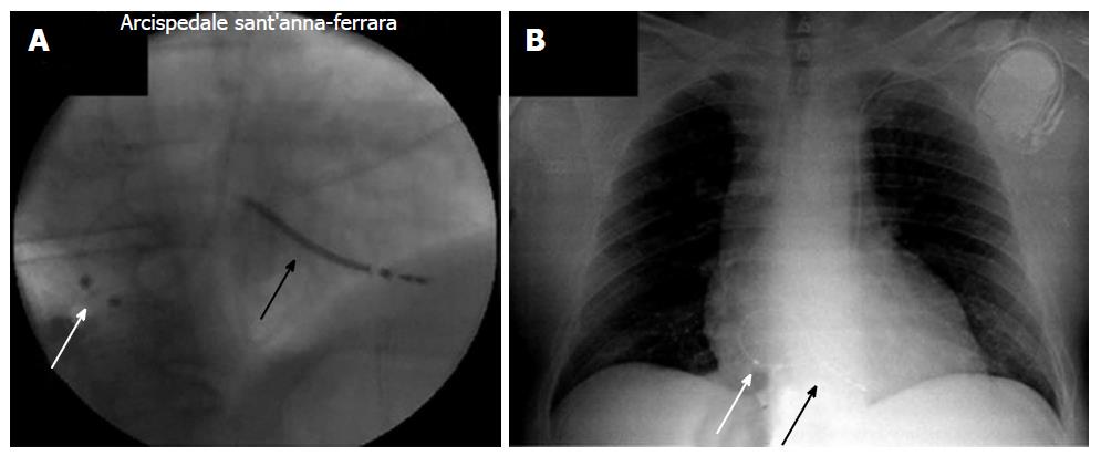
During the implant procedure, the catheter inserted via the left cephalic vein took an anomalous route to coronary sinus. A venous angiography via the cubitalis vein revealed a previously unknown persistence of LSVC draining into the coronary sinus. The right superior vena cava was present, normally draining into the right atrium. We then performed ICD DX implantation with insertion of a single-coil single lead with atrial sensing dipole (Biotronik Linox Smart S DX) via the left cephalic vein through LSVC and coronary sinus. The catheter was positioned in the right ventricular posterior wall towards the apex, with the atrial sensing dipole into the right atrium at a postero-inferior level and with the defibrillation coil near the interatrial septum inserted beyond the tricuspid valve (Figure 1). Electrical measurements showed acceptable values of atrial and ventricular sensing (4.3 mV and 5.7 mV, respectively), as well as ventricular pacing (0.6 V pacing threshold), impedance (377 Ohm) and shock impedance (65 Ohm). Total X-ray exposure time was 26 min and 24 s. Defibrillation test was not performed. The patient was then discharged and followed-up with remote monitoring (Biotronik Home Monitoring).
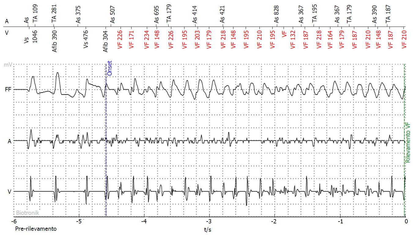
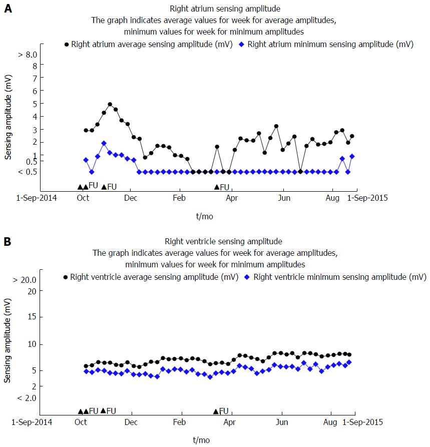
During 10 mo of follow-up, several events were reported. In particular, the patient experienced 4 episodes of sustained ventricular tachycardia/ventricular fibrillation. Of these, 3 were interrupted with antitachycardia pacing (ATP) and the fourth with a single 40 Joule DC-shock (vector between right ventricular coil and anterior can), which restored sinus rhythm (Figure 2). No atrial arrhythmias were detected. Diagnostics also revealed sensing/pacing time with 90% AS-VS, which indicate spontaneous rhythm, and only few times of pacing. Remote monitoring showed acceptable values of atrial and ventricular sensing, stable over time, indicating stable position of the lead (Figure 3).
In our patient, given the posterior position of RV catheter, we expect normal or even better efficacy of ICD since defibrillation vector, directed from posterior (right ventricle) to anterior (can) could include huge critical ventricular mass. This consideration should discourage the implantation in the right side. Furthermore, in one third of cases of LSVC there is absence of right superior vena cava[1]. Therefore, in patients with LSVC it is in any case more appropriate to implant the device on the left side.
In conclusion, this case report gives a contribution to the knowledge on this subject by confirming the possibility of successful implantation and effectiveness of the therapy. During 10 mo of follow-up, our patient presented a few episodes of ventricular arrhythmias, which were effectively recognized and treated either with ATP and with DC-shock.
Ischemic cardiomyopathy with reduced ejection fraction, scheduled for implantation of single lead implantable cardioverter defibrillator (ICD) with atrial sensing dipole.
Persistent left superior vena cava (LSVC).
Fluoroscopy and angiography during ICD implantation.
Single lead ICD with atrial sensing dipole implantation via persistent LSVC.
Stability of lead and effective ICD therapy both with antitachycardia pacing and DC-shock during follow-up.
Single lead ICD with atrial sensing dipole is a safe and effective technology even in patients with persistent LSVC.
The paper reports the implantation and follow-up data of a patient with persistent LSVC who underwent ICD implantation with a single lead capable of atrial sensing.
P- Reviewer: De Ponti R, Landesberg G, Petix NR S- Editor: Qi Y L- Editor: A E- Editor: Li D
| 1. | Biffi M, Bertini M, Ziacchi M, Martignani C, Valzania C, Diemberger I, Branzi A, Boriani G. Clinical implications of left superior vena cava persistence in candidates for pacemaker or cardioverter-defibrillator implantation. Heart Vessels. 2009;24:142-146. [RCA] [PubMed] [DOI] [Full Text] [Cited by in Crossref: 34] [Cited by in RCA: 35] [Article Influence: 2.2] [Reference Citation Analysis (0)] |
| 2. | Polewczyk A, Kutarski A, Czekajska-Chehab E, Adamczyk P, Boczar K, Polewczyk M, Janion M. Complications of permanent cardiac pacing in patients with persistent left superior vena cava. Cardiol J. 2014;21:128-137. [RCA] [PubMed] [DOI] [Full Text] [Cited by in Crossref: 14] [Cited by in RCA: 16] [Article Influence: 1.5] [Reference Citation Analysis (0)] |
| 3. | Guenther M, Kolschmann S, Rauwolf TP, Christoph M, Sandfort V, Strasser RH, Wunderlich C. Implantable cardioverter defibrillator lead implantation in patients with a persistent left superior vena cava--feasibility, chances, and limitations: representative cases in adults. Europace. 2013;15:273-277. [RCA] [PubMed] [DOI] [Full Text] [Cited by in Crossref: 12] [Cited by in RCA: 15] [Article Influence: 1.2] [Reference Citation Analysis (0)] |
| 4. | Morgan LG, Gardner J, Calkins J. The incidental finding of a persistent left superior vena cava: implications for primary care providers-case and review. Case Rep Med. 2015;2015:198754. [PubMed] |
| 5. | Iori M, Giacopelli D, Quartieri F, Bottoni N, Manari A. Implantable cardioverter defibrillator system with floating atrial sensing dipole: a single-center experience. Pacing Clin Electrophysiol. 2014;37:1265-1273. [RCA] [PubMed] [DOI] [Full Text] [Cited by in Crossref: 12] [Cited by in RCA: 16] [Article Influence: 1.5] [Reference Citation Analysis (0)] |
| 6. | Sun ZH, Stjernvall J, Laine P, Toivonen L. Extensive variation in the signal amplitude of the atrial floating VDD pacing electrode. Pacing Clin Electrophysiol. 1998;21:1760-1765. [RCA] [PubMed] [DOI] [Full Text] [Cited by in Crossref: 12] [Cited by in RCA: 14] [Article Influence: 0.5] [Reference Citation Analysis (0)] |


