
















©The Author(s) 2016.
World J Cardiol. Feb 26, 2016; 8(2): 132-145
Published online Feb 26, 2016. doi: 10.4330/wjc.v8.i2.132
Published online Feb 26, 2016. doi: 10.4330/wjc.v8.i2.132
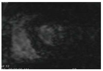
Figure 1 Iron overload cardiomyopathy.
Short axis gradient echo image with long echo time (15 ms) shows dark signal in the left ventricular myocardium due to increased iron deposition.
- Citation: Kalisz K, Rajiah P. Impact of cardiac magnetic resonance imaging in non-ischemic cardiomyopathies. World J Cardiol 2016; 8(2): 132-145
- URL: https://www.wjgnet.com/1949-8462/full/v8/i2/132.htm
- DOI: https://dx.doi.org/10.4330/wjc.v8.i2.132