Copyright
©The Author(s) 2016.
World J Cardiol. Dec 26, 2016; 8(12): 735-745
Published online Dec 26, 2016. doi: 10.4330/wjc.v8.i12.735
Published online Dec 26, 2016. doi: 10.4330/wjc.v8.i12.735
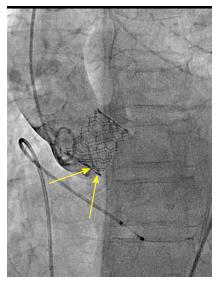
Figure 6 Example of difficult depth measurement.
In this case, the projection has been modified after implant so the device appears coaxial. However, the annulus is no longer coaxial: Two aortic cusps are seen at different levels on the septal side (arrows), making difficult the localization of the hinge point and therefore the measurement depth of implant.
- Citation: Sawaya FJ, Spaziano M, Lefèvre T, Roy A, Garot P, Hovasse T, Neylon A, Benamer H, Romano M, Unterseeh T, Morice MC, Chevalier B. Comparison between the SAPIEN S3 and the SAPIEN XT transcatheter heart valves: A single-center experience. World J Cardiol 2016; 8(12): 735-745
- URL: https://www.wjgnet.com/1949-8462/full/v8/i12/735.htm
- DOI: https://dx.doi.org/10.4330/wjc.v8.i12.735


