Copyright
©The Author(s) 2016.
World J Cardiol. Nov 26, 2016; 8(11): 667-675
Published online Nov 26, 2016. doi: 10.4330/wjc.v8.i11.667
Published online Nov 26, 2016. doi: 10.4330/wjc.v8.i11.667
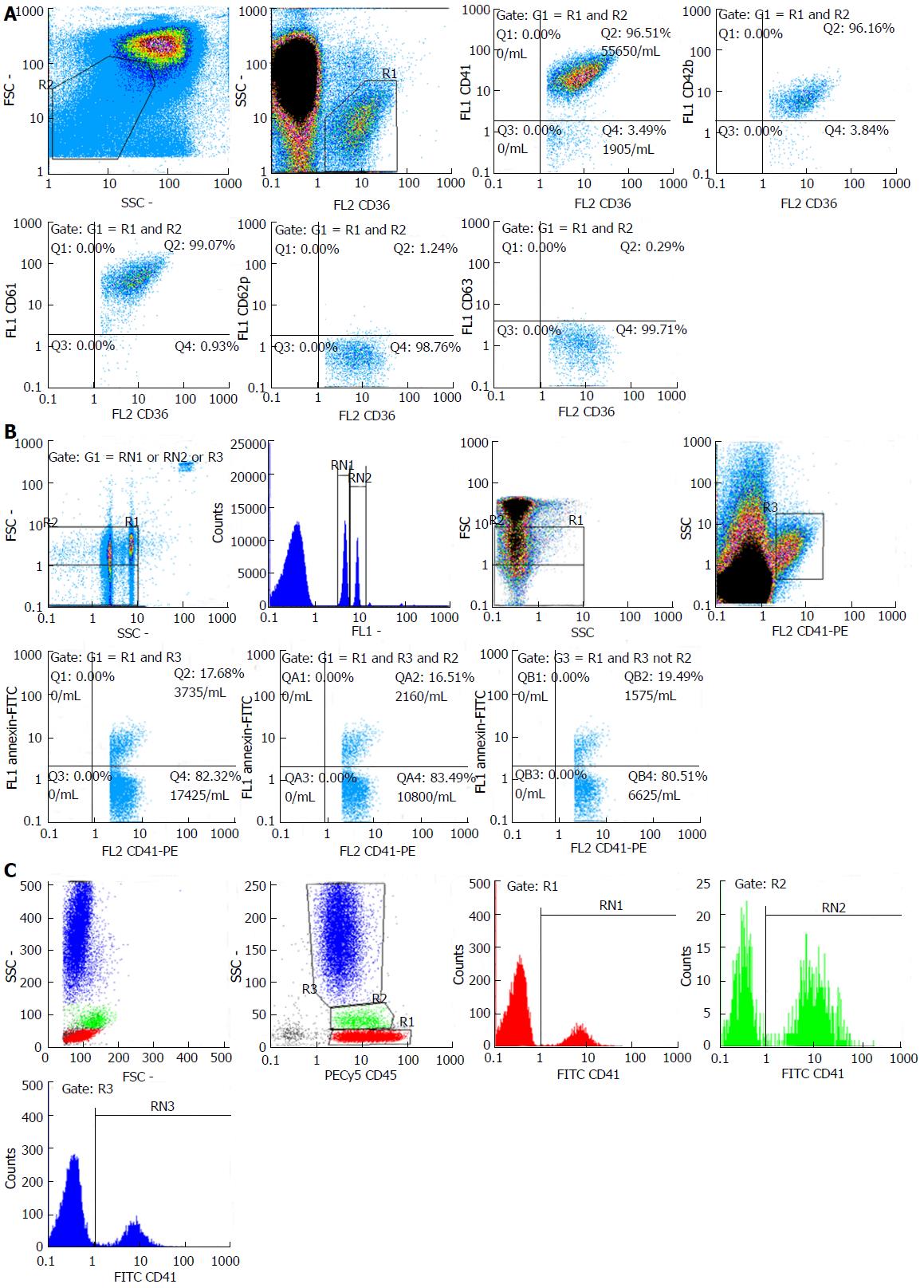
Figure 1 Flow cytometric analysis of platelet membrane glycoproteins, platelet-derived microparticles and platelet-leukocyte aggregates.
A: Flow cytometric analysis of membrane-bound glycoproteins. Analysis of each GP was performed on particles of FSC/SSC of platelets (R2) expressing CD36 (R1); B: Beads for gating of microparticles of 0.5 μm (left population), 0.9 μm (right population) and 3 μm (upper right population). Same gating strategy was used to identify PMPs of 0-0.5 μm (R1 and R3 not R2), 0.5-0.9 μm (R1 and R2 and R3) or PMPs in general (R1 and R3); C: Platelet-leukocyte aggregates calculated for lymphocytes (red), monocytes (green) and granulocytes (blue).
- Citation: Papadavid E, Diamanti K, Spathis A, Varoudi M, Andreadou I, Gravanis K, Theodoropoulos K, Karakitsos P, Lekakis J, Rigopoulos D, Ikonomidis I. Increased levels of circulating platelet-derived microparticles in psoriasis: Possible implications for the associated cardiovascular risk. World J Cardiol 2016; 8(11): 667-675
- URL: https://www.wjgnet.com/1949-8462/full/v8/i11/667.htm
- DOI: https://dx.doi.org/10.4330/wjc.v8.i11.667


