
















©The Author(s) 2015.
World J Cardiol. Jul 26, 2015; 7(7): 431-433
Published online Jul 26, 2015. doi: 10.4330/wjc.v7.i7.431
Published online Jul 26, 2015. doi: 10.4330/wjc.v7.i7.431
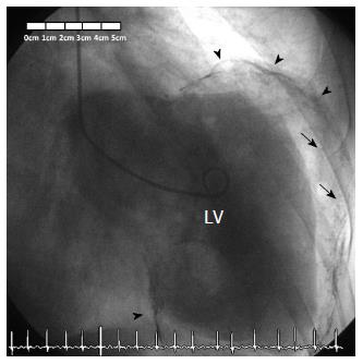
Figure 6 Left ventriculogram confirming diagnosis of a giant calcified and partially thrombosed left ventricular aneurysm, with severe left ventricular systolic dysfunction.
The wall of the aneurysm is calcified (arrowheads), and the aneurysm is covered with thrombus (arrows). LV: Left ventricle.
- Citation: de Agustin JA, de Diego JJG, Marcos-Alberca P, Rodrigo JL, Almeria C, Mahia P, Luaces M, Garcia-Fernandez MA, Macaya C, de Isla LP. Giant and thrombosed left ventricular aneurysm. World J Cardiol 2015; 7(7): 431-433
- URL: https://www.wjgnet.com/1949-8462/full/v7/i7/431.htm
- DOI: https://dx.doi.org/10.4330/wjc.v7.i7.431