
















©The Author(s) 2015.
World J Cardiol. Jul 26, 2015; 7(7): 415-422
Published online Jul 26, 2015. doi: 10.4330/wjc.v7.i7.415
Published online Jul 26, 2015. doi: 10.4330/wjc.v7.i7.415
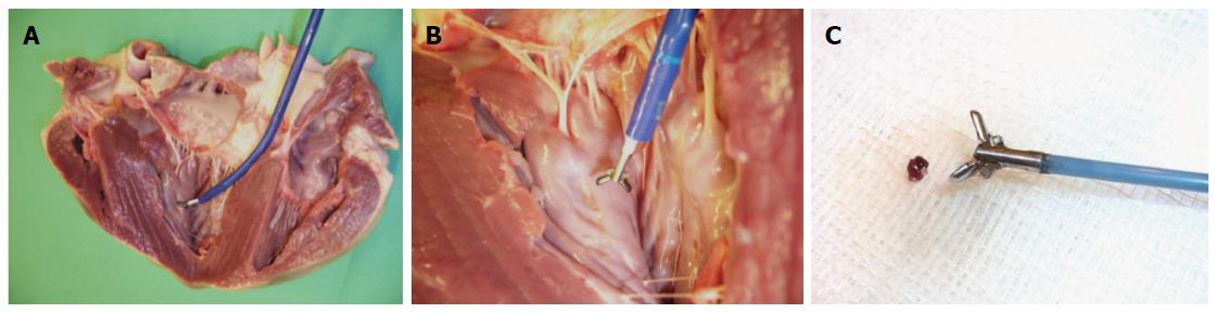
Figure 7 Close-up images of experimental in-vitro setup as shown in Figure 1: Opened porcine heart model with magnetic resonance bioptome (white) advanced through sheath (blue) (A), opened magnetic resonance bioptome forceps (B), magnetic resonance bioptome with tissue sample retrieved from endocardium (C).
-
Citation: Lossnitzer D, Seitz SA, Krautz B, Schnackenburg B, André F, Korosoglou G, Katus HA, Steen H. Feasibility of real-time magnetic resonance imaging-guided endomyocardial biopsies: An
in-vitro study. World J Cardiol 2015; 7(7): 415-422 - URL: https://www.wjgnet.com/1949-8462/full/v7/i7/415.htm
- DOI: https://dx.doi.org/10.4330/wjc.v7.i7.415