Copyright
©The Author(s) 2015.
World J Cardiol. Jun 26, 2015; 7(6): 361-366
Published online Jun 26, 2015. doi: 10.4330/wjc.v7.i6.361
Published online Jun 26, 2015. doi: 10.4330/wjc.v7.i6.361
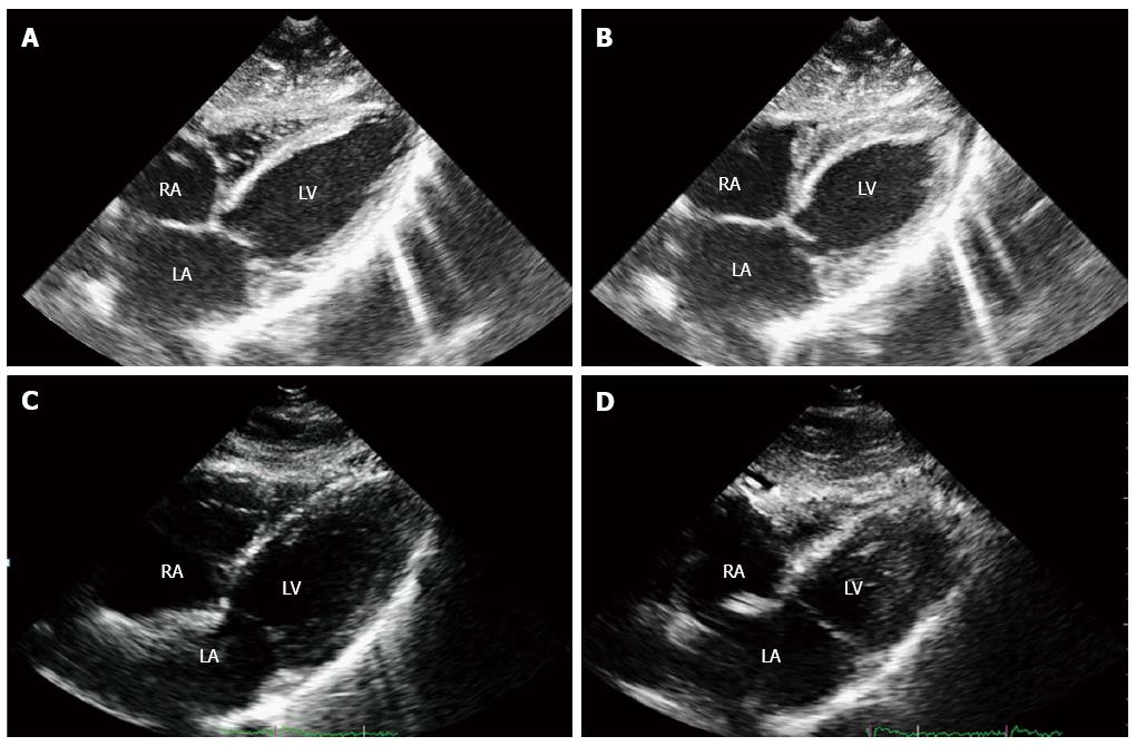
Figure 3 Two-dimensional echocardiogram, subcostal four chambers view, showing the anteroseptal and posterolateral walls of the left ventricle.
End-diastolic (A) and mid-systolic (B) frames at the time of acute cardiac symptoms presentation showed dyskinesia of basal and medium segments, with hyperkinesia of the left ventricular apex. One week later, recovery of the wall motion abnormalities was demonstrated, with hyperdynamic ejection fraction (C and D). A previous echocardiogram performed two years before in this patient was similar to this last one. RA: Right atrium; LA: Left atrium; LV: Left ventricle.
- Citation: Robles P, Monedero I, Rubio A, Botas J. Reverse or inverted apical ballooning in a case of refeeding syndrome. World J Cardiol 2015; 7(6): 361-366
- URL: https://www.wjgnet.com/1949-8462/full/v7/i6/361.htm
- DOI: https://dx.doi.org/10.4330/wjc.v7.i6.361


