Copyright
©The Author(s) 2015.
World J Cardiol. Feb 26, 2015; 7(2): 104-110
Published online Feb 26, 2015. doi: 10.4330/wjc.v7.i2.104
Published online Feb 26, 2015. doi: 10.4330/wjc.v7.i2.104
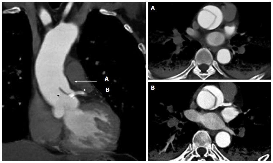
Figure 5 Contrast-enhanced computed tomography after coronary stenting.
The sagittal view (left image) reveals the localized dissection of the ascending aorta that extends to the left main coronary artery ostium. The implanted coronary stent (arrow) protects against the intrusion of the dissection flap (black arrowhead) into the left coronary artery. Ascending aortic dissection is clearly visible in the horizontal view (right images) in accordance with A and B in the sagittal view.
- Citation: Hanaki Y, Yumoto K, I S, Aoki H, Fukuzawa T, Watanabe T, Kato K. Coronary stenting with cardiogenic shock due to acute ascending aortic dissection. World J Cardiol 2015; 7(2): 104-110
- URL: https://www.wjgnet.com/1949-8462/full/v7/i2/104.htm
- DOI: https://dx.doi.org/10.4330/wjc.v7.i2.104


