Copyright
©The Author(s) 2015.
World J Cardiol. Nov 26, 2015; 7(11): 776-783
Published online Nov 26, 2015. doi: 10.4330/wjc.v7.i11.776
Published online Nov 26, 2015. doi: 10.4330/wjc.v7.i11.776
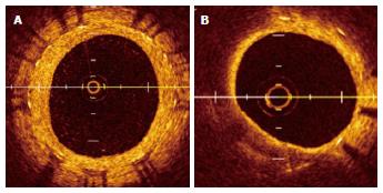
Figure 2 Optical coherence tomography images of common neointima (A) and neoatherosclerosis (B).
A: Common neointima is recognized by its high-signal intensity and homogeneous region inside stent struts; B: The neointima has a diffuse border and marked attenuation.
- Citation: Komiyama H, Takano M, Hata N, Seino Y, Shimizu W, Mizuno K. Neoatherosclerosis: Coronary stents seal atherosclerotic lesions but result in making a new problem of atherosclerosis. World J Cardiol 2015; 7(11): 776-783
- URL: https://www.wjgnet.com/1949-8462/full/v7/i11/776.htm
- DOI: https://dx.doi.org/10.4330/wjc.v7.i11.776


