Copyright
©2014 Baishideng Publishing Group Co.
World J Cardiol. Apr 26, 2014; 6(4): 196-204
Published online Apr 26, 2014. doi: 10.4330/wjc.v6.i4.196
Published online Apr 26, 2014. doi: 10.4330/wjc.v6.i4.196
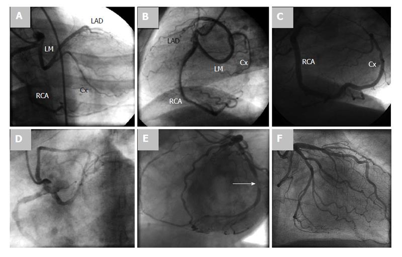
Figure 2 Coronary angiography frame.
A: Coronary angiography frame of right anterior oblique projection with cranial angulation; B: Left lateral (LL) projection showing a single origin of the right and left coronary arteries from a common right coronary ostium (Lipton R-IIP), the long curved left main stem and right dominancy are delineated; C: Coronary angiography frame in LL projection demonstrating a single coronary artery originating from the right sinus of Valsalva (RSV) giving the left anterior descending (LAD) and continued as the circumflex artery (Lipton R-I); D: Coronary angiography frame in left anterior oblique view demonstrating a single coronary artery arising from RSV as a single unique ostium (Lipton R-III); E: Coronary angiography frame in left anterior oblique view showing a single coronary artery originating from the left sinus of Valsalva. The terminal branch of circumflex artery represented the right coronary artery (Lipton L-I). Significant stenosis of the mid circumflex artery is demonstrated (white arrow); F: Coronary angiography frame demonstrates appearance of both right and left coronary arteries on injection of left sinus of Valsalva, as a single common ostium (Lipton L-IIA). Cx: Circumflex artery; RCA: Right coronary artery.
- Citation: Said SA, de Voogt WG, Bulut S, Han J, Polak P, Nijhuis RL, op den Akker JW, Slootweg A. Coronary artery disease in congenital single coronary artery in adults: A Dutch case series. World J Cardiol 2014; 6(4): 196-204
- URL: https://www.wjgnet.com/1949-8462/full/v6/i4/196.htm
- DOI: https://dx.doi.org/10.4330/wjc.v6.i4.196


