Copyright
©2014 Baishideng Publishing Group Co.
World J Cardiol. Apr 26, 2014; 6(4): 183-195
Published online Apr 26, 2014. doi: 10.4330/wjc.v6.i4.183
Published online Apr 26, 2014. doi: 10.4330/wjc.v6.i4.183
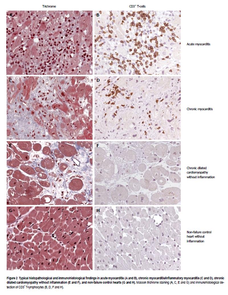
Figure 2 Typical histopathological and immunohistological findings in acute myocarditis (A and B), chronic myocarditis/inflammatory myocarditis (C and D), chronic dilated cardiomyopathy without inflammation (E and F), and non-failure control hearts (G and H).
Masson trichrome staining (A, C, E and G) and immunohistological detection of CD3+ T-lymphocytes (B, D, F and H).
- Citation: Bock CT, Düchting A, Utta F, Brunner E, Sy BT, Klingel K, Lang F, Gawaz M, Felix SB, Kandolf R. Molecular phenotypes of human parvovirus B19 in patients with myocarditis. World J Cardiol 2014; 6(4): 183-195
- URL: https://www.wjgnet.com/1949-8462/full/v6/i4/183.htm
- DOI: https://dx.doi.org/10.4330/wjc.v6.i4.183


