Copyright
©2014 Baishideng Publishing Group Inc.
World J Cardiol. Dec 26, 2014; 6(12): 1234-1244
Published online Dec 26, 2014. doi: 10.4330/wjc.v6.i12.1234
Published online Dec 26, 2014. doi: 10.4330/wjc.v6.i12.1234
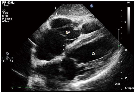
Figure 5 Two-dimensional echocardiogram, 4 chamber subcostal view, end diastolic frame, in an arrhythmogenic right ventricular cardiomyopathy patient.
RV wall aneurysm at subtricuspid basal level is evident (arrow). LV: Left ventricle; RV: Right ventricle.
- Citation: Pinamonti B, Brun F, Mestroni L, Sinagra G. Arrhythmogenic right ventricular cardiomyopathy: From genetics to diagnostic and therapeutic challenges. World J Cardiol 2014; 6(12): 1234-1244
- URL: https://www.wjgnet.com/1949-8462/full/v6/i12/1234.htm
- DOI: https://dx.doi.org/10.4330/wjc.v6.i12.1234


