Copyright
©2014 Baishideng Publishing Group Inc.
World J Cardiol. Nov 26, 2014; 6(11): 1223-1226
Published online Nov 26, 2014. doi: 10.4330/wjc.v6.i11.1223
Published online Nov 26, 2014. doi: 10.4330/wjc.v6.i11.1223
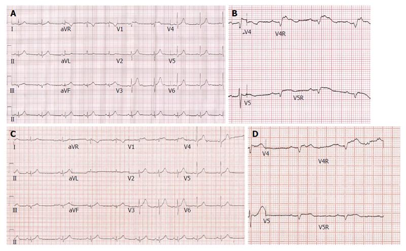
Figure 1 Electrocardiography.
A: Standard 12-lead ECG at time of presentation showing ST elevation in lead V1; B: Right-sided ECG leads showing ST elevation in V4R and V5R; C: 12-lead ECG post-primary percutaneous intervention showing partial resolution of ST elevation in V1; D: Right-sided ECG post- percutaneous intervention showing resolution of ST elevation in V4R and V5R. ECG: Electrocardiography.
- Citation: Chahal AA, Kim MY, Borg AN, Al-Najjar Y. Primary angioplasty for infarction due to isolated right ventricular artery occlusion. World J Cardiol 2014; 6(11): 1223-1226
- URL: https://www.wjgnet.com/1949-8462/full/v6/i11/1223.htm
- DOI: https://dx.doi.org/10.4330/wjc.v6.i11.1223


