Copyright
©2014 Baishideng Publishing Group Inc.
World J Cardiol. Oct 26, 2014; 6(10): 1127-1130
Published online Oct 26, 2014. doi: 10.4330/wjc.v6.i10.1127
Published online Oct 26, 2014. doi: 10.4330/wjc.v6.i10.1127
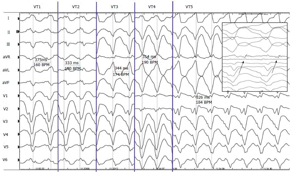
Figure 3 Cycle and QRS morphologies of all the 5 sustained ventricular tachyarrhythmias observed and mapped during the procedure.
In the box it is possible to appreciate the mid-diastolic potential (arrows) recorded during ventricular tachyarrhythmias (VT) 5. BPM: Beat per minute.
- Citation: Casella M, Carbucicchio C, Russo E, Pizzamiglio F, Golia P, Conti S, Costa F, Russo AD, Tondo C. Electrical storm in systemic sclerosis: Inside the electroanatomic substrate. World J Cardiol 2014; 6(10): 1127-1130
- URL: https://www.wjgnet.com/1949-8462/full/v6/i10/1127.htm
- DOI: https://dx.doi.org/10.4330/wjc.v6.i10.1127


