Copyright
©2013 Baishideng Publishing Group Co.
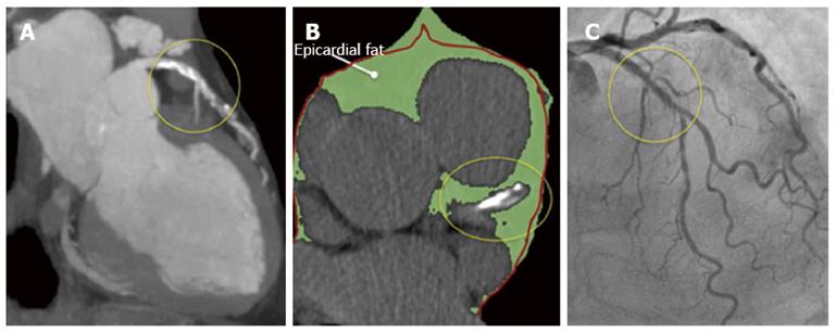
Figure 2 A multislice computed tomography was performed in a 68-year old male because of atypical chest pain.
A: This study revealed a long mixed, calcific and non-calcific plaque in the proximal and mid segments of the left anterior descending artery; B: A large amount of epicardial adipose tissue (EAT) [(159.2 cm3 (green) (figure 3)] was also calculated. Please note that the left anterior descending artery is embedded in EAT (yellow circles). The red line represents the pericardium; C: A coronary angiogram was scheduled and revealed intermediate stenoses in the same arterial segment that was subsequently studied with optical coherence tomography.
- Citation: Echavarría-Pinto M, Hernando L, Alfonso F. From the epicardial adipose tissue to vulnerable coronary plaques. World J Cardiol 2013; 5(4): 68-74
- URL: https://www.wjgnet.com/1949-8462/full/v5/i4/68.htm
- DOI: https://dx.doi.org/10.4330/wjc.v5.i4.68


