
















©2012 Baishideng Publishing Group Co.
World J Cardiol. Oct 26, 2012; 4(10): 284-287
Published online Oct 26, 2012. doi: 10.4330/wjc.v4.i10.284
Published online Oct 26, 2012. doi: 10.4330/wjc.v4.i10.284
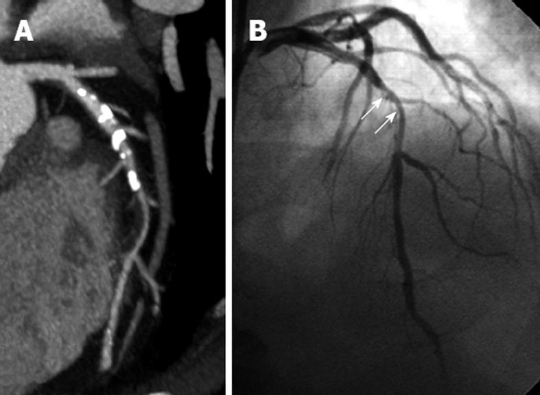
Figure 2 Calcified plaques and stenosis of left anterior descending.
A: Extensive calcified plaques are noticed in the proximal and middle segments of left anterior descending (LAD) on curved planar reformatted image, resulting in significant stenosis or total lumen occlusion; B: A 50% stenosis of LAD is confirmed on invasive coronary angiography (arrows).
- Citation: Almoudi M, Sun Z. Coronary artery calcium score: Re-evaluation of its predictive value for coronary artery disease. World J Cardiol 2012; 4(10): 284-287
- URL: https://www.wjgnet.com/1949-8462/full/v4/i10/284.htm
- DOI: https://dx.doi.org/10.4330/wjc.v4.i10.284