Copyright
©2011 Baishideng Publishing Group Co.
World J Cardiol. May 26, 2011; 3(5): 127-134
Published online May 26, 2011. doi: 10.4330/wjc.v3.i5.127
Published online May 26, 2011. doi: 10.4330/wjc.v3.i5.127
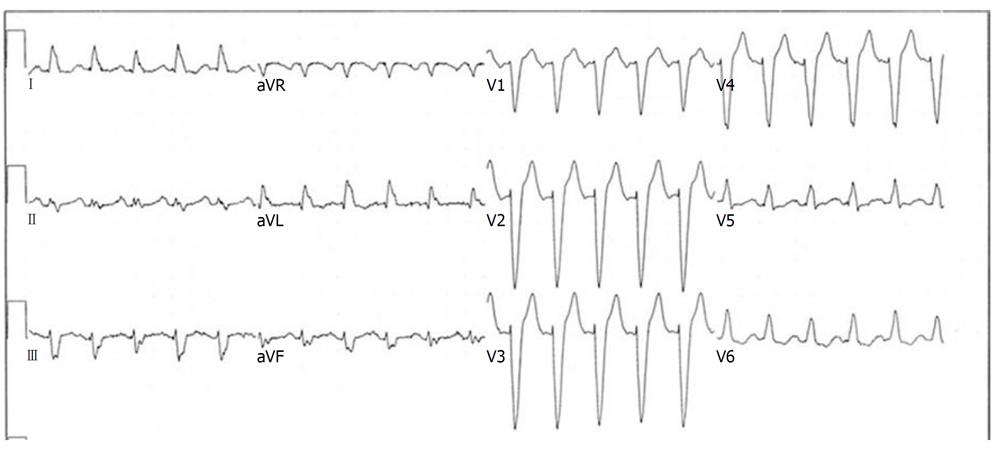
Figure 1 Electrocardiogram example of typical left bundle branch block pattern in a patient with sinus tachycardia.
Lead V1 demonstrates an rS complex, while there are monophasic, notched R waves in leads I, aVL, V5 and V6. Q waves are absent in these leads.
- Citation: Neiger JS, Trohman RG. Differential diagnosis of tachycardia with a typical left bundle branch block morphology. World J Cardiol 2011; 3(5): 127-134
- URL: https://www.wjgnet.com/1949-8462/full/v3/i5/127.htm
- DOI: https://dx.doi.org/10.4330/wjc.v3.i5.127


