Copyright
©2010 Baishideng Publishing Group Co.
World J Cardiol. Oct 26, 2010; 2(10): 344-356
Published online Oct 26, 2010. doi: 10.4330/wjc.v2.i10.344
Published online Oct 26, 2010. doi: 10.4330/wjc.v2.i10.344
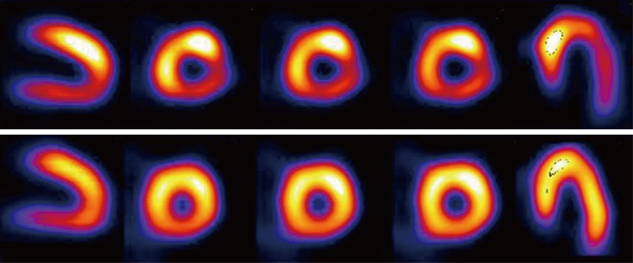
Figure 1 Single photon emission computed tomography image of exercise 201Tl scintigraphy in a 70-year-old man.
The stress image (upper panel) shows decreased perfusion in the infero-lateral region. There is a redistribution of the tracer in the rest image (lower panel), which indicates exercise-induced myocardial ischemia in the infero-lateral region of the left ventricle.
- Citation: Matsuo S, Nakajima K, Kinuya S. Clinical use of nuclear cardiology in the assessment of heart failure. World J Cardiol 2010; 2(10): 344-356
- URL: https://www.wjgnet.com/1949-8462/full/v2/i10/344.htm
- DOI: https://dx.doi.org/10.4330/wjc.v2.i10.344


