
















©2010 Baishideng Publishing Group Co.
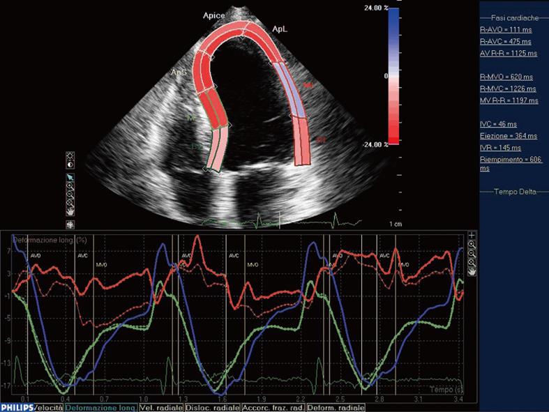
Figure 2 Comparison between epicardial (continous line) and endocardial border (discontinuous line) in lateral myocardial infarction.
In the mid-septal segment (green line) the endocardial and epicardial curves are indistinguishable and result in an end-systolic shortening, while in the ischemic mid-lateral segment (red line) the epicardial curve separates from the endocardial one. Moreover, in this segment, we can observe the lack of myocardial deformation after the electromechanical activation.
- Citation: Sitia S, Tomasoni L, Turiel M. Speckle tracking echocardiography: A new approach to myocardial function. World J Cardiol 2010; 2(1): 1-5
- URL: https://www.wjgnet.com/1949-8462/full/v2/i1/1.htm
- DOI: https://dx.doi.org/10.4330/wjc.v2.i1.1