
















Copyright: ©Author(s) 2026.
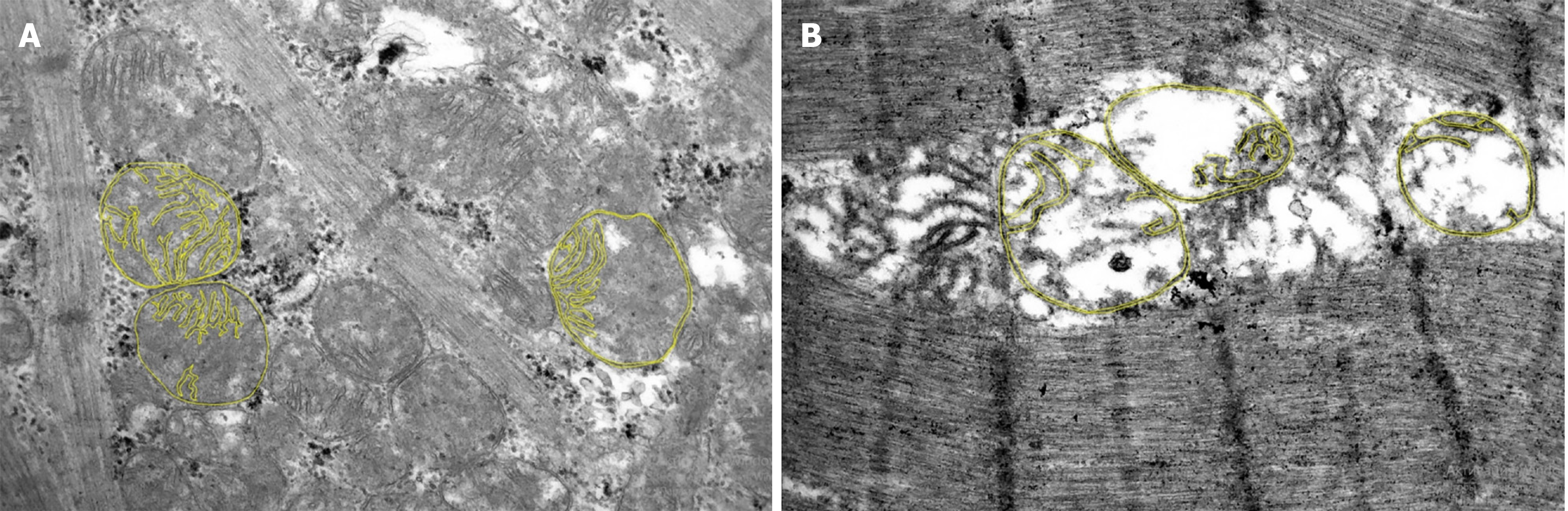
Figure 5 Micrographs of a cardiomyocyte from the auricle of the right atrium in two patients with ischemic heart failure (magnification × 15000).
Yellow lines indicate the outer and inner membranes of three mitochondria for each patient to calculate the “Ratio of mitochondrial membrane lengths”. A: Micrograph of the myocardium of a surviving patient, where the “Ratio of mitochondrial membrane lengths” was 21%; B: Micrograph of the myocardium of a deceased patient, where the “Ratio of mitochondrial membrane lengths” was 53% (a predictor of mortality).
- Citation: Kuzheleva EA, Garganeeva AA, Tukish OV, Syromyatnikova EE. Mitochondrial ultrastructure in cardiac myocytes is associated with the cardiovascular death in patients with heart failure. World J Cardiol 2026; 18(8): 124556
- URL: https://www.wjgnet.com/1949-8462/full/v18/i8/124556.htm
- DOI: https://dx.doi.org/10.4330/wjc.124556