
















Copyright: ©Author(s) 2026.
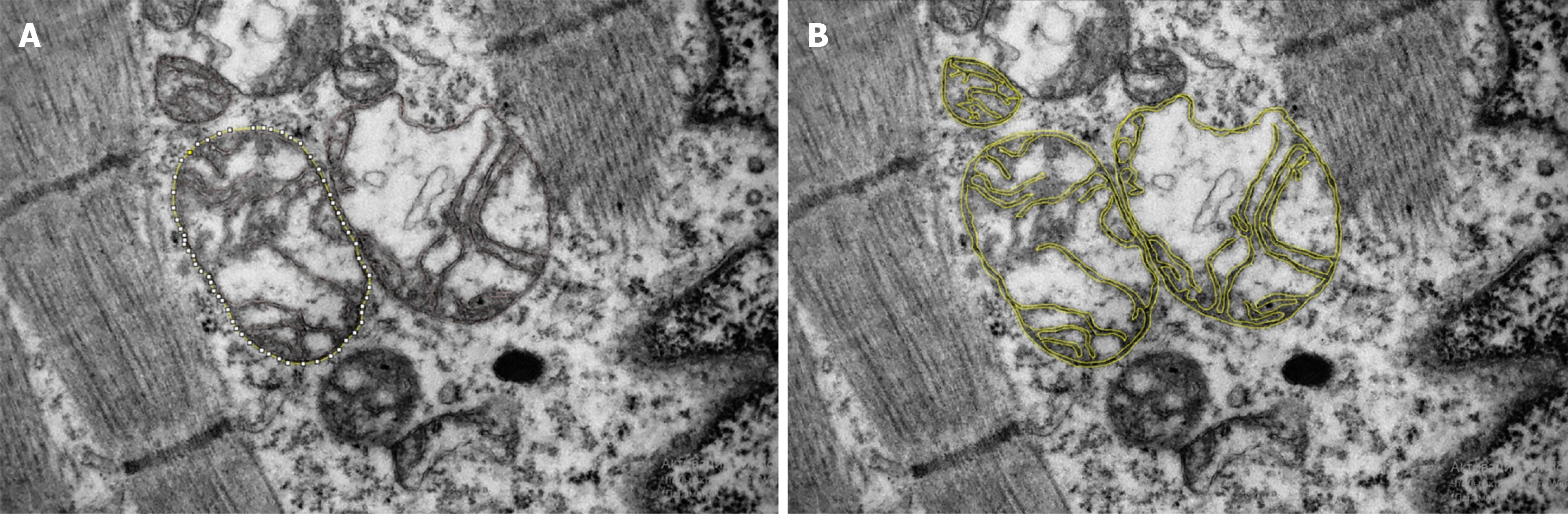
Figure 3 Micrograph of a cardiomyocyte from the auricle of the right atrium in a patient with ischemic heart failure (magnification × 15000).
An example of the separation of the inner and outer mitochondrial membranes is shown in the micrograph (yellow line), which can be used to calculate the ratio of the outer to inner mitochondrial membrane length. A: A yellow line outlines the outer membrane of a single mitochondrion; B: Yellow lines indicate the outer and inner membranes of three mitochondria.
- Citation: Kuzheleva EA, Garganeeva AA, Tukish OV, Syromyatnikova EE. Mitochondrial ultrastructure in cardiac myocytes is associated with the cardiovascular death in patients with heart failure. World J Cardiol 2026; 18(8): 124556
- URL: https://www.wjgnet.com/1949-8462/full/v18/i8/124556.htm
- DOI: https://dx.doi.org/10.4330/wjc.124556