
















Copyright: ©Author(s) 2026.
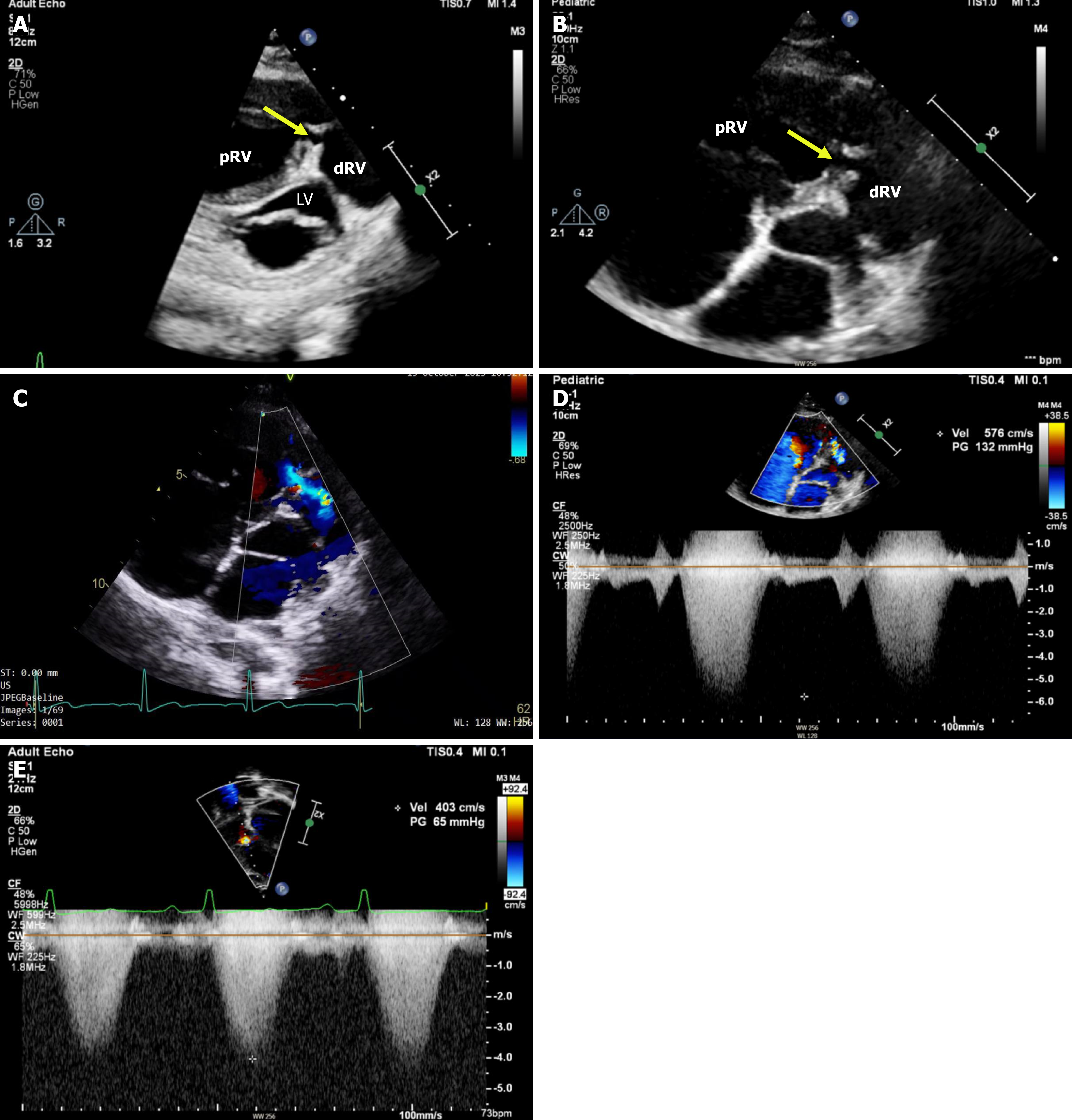
Figure 2 Echocardiographic features of double-chambered right ventricle with right ventricular obstruction and Doppler assessment.
A: Trans-thoracic echocardiogram, parasternal modified short-axis view (at mitral valve level) demonstrating a double-chambered right ventricle with a tunnel-like obstruction (arrow) due to a prominent right ventricular muscle bundle and right ventricular hypertrophy. The arrow highlights a right ventricular muscle bundle that forms the tunnel-like obstruction; B: Trans-thoracic echocardiogram, parasternal modified short-axis view demonstrating a double-chambered right ventricle with a tunnel-like obstruction (arrow) due to a prominent right ventricular muscle bundle. The arrow highlights a right ventricular muscle bundle that forms the tunnel-like obstruction; C: Color-Doppler demonstrating turbulence of flow across the right ventricular muscle bundle; D: Continuous-wave Doppler across the area of obstruction (intra-cavity), with an estimated peak gradient of 132 mmHg; E: Continuous-wave Doppler across the tricuspid valve, with an estimated peak gradient of 65 mmHg. dRV: Distal right ventricular chamber; LV: Left ventricular cavity; pRV: Proximal right ventricular chamber.
- Citation: Alahmed YS, Almedaihesh RF, Alshammari HA, Alrusayni O, Alhawri KA. Isolated double-chambered right ventricle in an adult: A case report. World J Cardiol 2026; 18(8): 123370
- URL: https://www.wjgnet.com/1949-8462/full/v18/i8/123370.htm
- DOI: https://dx.doi.org/10.4330/wjc.123370